REVIEW ARTICLE


Primary Hyperparathyroidism in the Common Orthopaedic Practice


Nikolaos K. Sferopoulos1, *
1 2nd Department of Orthopaedic Surgery, Aristotle University of Thessaloniki, “G. Gennimatas” Hospital, 546 35, Thessaloniki, Greece


Article Metrics

CrossRef Citations:
0
Total Statistics:

Full-Text HTML Views: 1777
Abstract HTML Views: 395
PDF Downloads: 333
ePub Downloads: 220
Total Views/Downloads: 2725
Unique Statistics:

Full-Text HTML Views: 822
Abstract HTML Views: 262
PDF Downloads: 264
ePub Downloads: 173
Total Views/Downloads: 1521


Creative Commons License
© 2021 Nikolaos K Sferopoulos

open-access license: This is an open access article distributed under the terms of the Creative Commons Attribution 4.0 International Public License (CC-BY 4.0), a copy of which is available at: https://creativecommons.org/licenses/by/4.0/legalcode. This license permits unrestricted use, distribution, and reproduction in any medium, provided the original author and source are credited.

* Address correspondence to this author at the 2nd Department of Orthopaedic Surgery, Aristotle University of Thessaloniki, “G. Gennimatas” Hospital, 546 35, Thessaloniki, Greece; Tel: 00302310963270, Fax: 00302310968265; E-mail: sferopoulos@yahoo.com


Abstract

An extensive review of the publications on primary hyperparathyroidism (pHPT) is presented in this report. It has strongly been emphasized in the literature that patients with pHPT may present either with the classical symptomatology or with asymptomatic disease, emerged due to biochemical screening. The clinical and epidemiological presentation of pHPT in western countries has changed profoundly during the past few decades, and bone disease is nowadays a distinct rarity. The introduction of serum calcium screening for osteoporosis and the technological advances in the laboratory assessment of parathyroid hormone have played important roles in early diagnosis. Subsequently, the disease is increasingly being detected as asymptomatic hypercalcaemia without guiding signs or symptoms. A third type of disease, the normocalcaemic variant, has been recently described in the literature. However, the potential diagnosis of pHPT should always be on the orthopaedics’ list of differential diagnoses in female or elderly patients with vertebral fractures and nephrolithiasis, either symptomatic or asymptomatic, as well as when solitary or multiple osteolytic lesions are encountered on the radiographs.

Additionally, a middle aged woman with parathyroid adenoma and subsequent brown tumors detected on the pelvic radiographs is reported. Her initial laboratory findings indicated a minimal increase of the serum calcium, a mild increase of the erythrocyte sedimentation rate, and a significant increase in total serum alkaline phosphatase. Finally, the detection of elevated parathyroid hormone levels indicated the diagnosis of pHPT and necessitated imaging studies of the parathyroid glands, which indicated a parathyroid adenoma. Following successful excision of the parathyroid adenoma, the patient suffered from the hungry bone syndrome. After a follow-up of 20 years, the patient had normal calcium, vitamin D, and parathyroid hormone serum levels, while a pelvic radiograph indicated no significant changes in the appearance of the brown tumors.

Keywords: Primary hyperparathyroidism, Parathyroid hormone, Hypercalcaemia, Vitamin D, Osteoporosis, Brown tumor, Hungry bone syndrome.


1. INTRODUCTION

The purpose of this report is to perform an extensive review of the relevant international literature about primary hyperparathyroidism (pHPT), to indicate the value of clinical suspicion for the early diagnosis of the disease in a patient presenting in the common orthopaedic practice, and to present an illustrative case with a 20-year follow-up.

2. REVIEW

2.1. Parathyroid Hormone

The parathyroid hormone, also called parathormone (PTH), is secreted by the chief cells of the four parathyroid glands. It is a cornerstone of calcium homeostasis through its increased calcium absorption effect in the gastrointestinal tract, kidney, and the skeleton by osteoclasts. It also plays a role in activating vitamin D. It is a polypeptide containing 84 amino acids, whose action is opposed by the hormone calcitonin. The PTH has both anabolic and catabolic skeletal effects that vary according to the kinetics of serum levels and the type of bone. The anabolic effects are manifested in patients with a periodic, rapid, and transient rise in serum PTH, as seen with the daily subcutaneous injection of recombinant human PTH (1-34) and PTH (1-84) in the treatment of osteoporosis. These patients have increased Bone Mineral Density (BMD) at skeletal sites with a high trabecular component, such as the lumbar spine, and a reduction in fracture risk. The catabolic effects are typified in patients with chronic, persistently elevated PTH levels.

2.2. Hyperparathyroidism

Hyperparathyroidism (HPT) is due to increased activity of the parathyroid glands either from an intrinsic abnormal change altering excretion of PTH (primary and tertiary HPT) or from an extrinsic abnormal change affecting calcium homeostasis stimulating the production of PTH (secondary HPT). Secondary HPT (sHPT) may result from a primary response to a physiological stimulus, such as in calcium malabsorption syndromes, chronic renal disease, or chronic hypocalcaemia following vitamin D deficiency, particularly in elderly people. Hyperparathyroidism may persist in a significant proportion of patients with sHPT and chronic kidney disease after kidney transplantation, a state known as tertiary HPT.

All types of HPT have many symptomatic features and skeletal manifestations in common. From a biochemical perspective, their main difference is that pHPT causes an increase in serum calcium and reduces phosphate, while sHPT produces the opposite [1-4].

2.2.1. Primary Hyperparathyroidism

2.2.1.1. Introduction

Primary hyperparathyroidism is one of the most common endocrine diseases. It is characterized by the autonomous, incompletely regulated, excessive secretion of PTH. It is most commonly sporadic. It is caused by a solitary parathyroid gland adenoma, usually affecting a single parathyroid gland, in 80-85% of the patients, and in 15-20%, it is associated with all four-gland parathyroid hyperplasia or multiple adenomas in hereditary or familial syndromes, while less than 1% is due to carcinoma. The female to male ratio is approximately 3:1. It is usually diagnosed in postmenopausal women, although younger patients and women entering menopause may also be involved. Three forms of the disease have been described so far. It may appear with the typical symptomatology or with atypical clinical findings, while a third type, the normocalcaemic variant, has recently been discovered. It is the commonest cause of hypercalcaemia (excluding humoral hypercalcaemia of malignancy). The disease is traditionally diagnosed by hypercalcaemia associated with elevated PTH serum levels, a critical finding for the differentiation of pHPT from diseases or tumors associated with hypercalcaemia. Hypercalcaemia is defined as raised total and ionized serum calcium concentrations; severe hypercalcaemia is associated with acute signs and symptoms, while mild hypercalcaemia is often asymptomatic. Normocalcaemic pHPT is an uncommon condition, a forme fruste of the disease. It is defined by normal total and ionized serum calcium concentrations but with PTH levels that are consistently elevated in the absence of secondary causes of HPT. The elevated PTH should be detected by the more recent assays that detect the 1-84 full-length molecule [5-24].

2.2.1.2. Clinical Presentation

Several studies have suggested that pHPT may be associated not only with the classical renal stones and bone manifestations but also with non-classical ones, such as cardiovascular, gastrointestinal, myotendinous, articular and neuropsychological symptoms, impaired quality of life, and increased cancer risk. Patients usually present with systemic symptoms of HPT or local clinical findings of related skeletal lesions. General early symptoms usually include fatigue, weight loss, difficulty in concentrating, somnolence, muscle weakness, hyperreflexia, and polyuria-polydipsia. Patients affected by pHPT are often misdiagnosed because their dominant picture may include nonspecific symptoms due to severe hypercalcaemia, such as gastritis, constipation, and depression, leading to diagnostic confusion. In severe symptomatic pHPT, renal involvement is manifested by hypercalciuria, nephrolithiasis, and nephrocalcinosis. The disease is also cited as one of the causal diseases that induce secondary osteoporosis. Osteoporosis and hypercalciuria associated with vertebral fractures and nephrolithiasis, respectively, may occasionally be asymptomatic. Nonetheless, the routine use of imaging and of biochemical determinations have revealed the frequent occurrence of asymptomatic reduced kidney function, in terms of estimated glomerular filtration rate, as well as increased morbidity and mortality, which has been defined as chronic kidney disease. In elderly patients, pHPT is more frequently asymptomatic and mostly characterized by bone involvement, while younger patients are more often symptomatic and mainly affected by renal impairment [25-36]. Data from the international literature have revealed the increased correlation of urine calcium excretion with the risk of stone formation. Moreover, calcium excretion is significantly greater in younger patients with pHPT [37-42]. Patients with normocalcaemic pHPT have more substantial skeletal involvement than is typical, and they develop more findings and complications over time [43]. It has been emphasized in the world publications that pHPT should always be included in the differential diagnosis of patients with a history of nephrolithiasis, nephrocalcinosis, osseous pain, osteoporosis-osteopenia, and subperiosteal bone resorption, as well as in patients with a personal history of neck irradiation, lithium therapy or a family history of multiple endocrine neoplasia syndrome [4, 44]. Recent observations have suggested an association between vitamin D deficiency and severity of pHPT, indicating that the disease seems to be more severe in those with concomitant vitamin D deficiency, while vitamin D deficiency and insufficiency seem to be more prevalent in patients with pHPT than in geographically matched populations [45-50].

During the past few decades, the clinical presentation of pHPT has changed globally towards an oligo- or asymptomatic disease with uncertain and long-lasting progression, while most patients have minimal overt symptomatology with no renal or skeletal manifestations. Since the frequent, routinely performed measurement evaluation of serum calcium levels in developed countries using automated multichannel analyzers and the development of assays for intact 1-84PTH, the diagnosis is usually made during osteoporosis screening or fortuitously in asymptomatic patients. Many publications have shown that the incidence increases with age and is higher in women [51-54]. In contrast, paediatric pHPT is considered a very rare endocrinopathy. It is frequently associated with pathological fractures; therefore, it may be easily misdiagnosed as osteogenesis imperfecta [55, 56].

2.2.1.3. Bone Disease

The chronic excessive hypersecretion of PTH in pHPT has a significant impact on bone remodeling. Parathyroid hormone stimulates both bone resorption and bone formation. Markers of the increased bone turnover include bone formation markers (serum alkaline phosphatase, serum bone alkaline phosphatase, and serum osteocalcin), bone resorption markers, and longitudinal bone turnover marker studies. Alkaline phosphatase level, the most widely clinically available marker, can be mildly elevated but in many patients is within normal limits. In addition, bone densitometry, bone histomorphometry, and newer non-invasive imaging techniques offer insights into the skeletal manifestations of pHPT. Bone turnover increases by about 50%, leading to increased resorption at the endosteal envelope, increased cortical porosity, and thinning of cortical bone with a decrease in BMD. Bone density, as measured by photon absorptiometry and bone histomorphometry, shows a deficit of cortical bone and preservation or increase in cancellous bone elements in mild pHPT with no clinical evidence of skeletal disease. Bone loss may also be readily appreciated by densitometry, showing a typical pattern in which cancellous bone of the lumbar spine is reasonably well preserved, whilst the cortical bone of the distal third of the radius is preferentially reduced. The latter is a relatively uniform finding and is associated with an increase in forearm fracture risk. Bone densitometry is advised, particularly for monitoring bone mass at the midradius or femoral neck, in patients with pHPT. Most densitometry studies support the concept that the PTH appears to be catabolic at cortical sites and may have anabolic effects at cancellous bone sites [57-64]. However, some studies have indicated that the overall fracture risk is increased in untreated pHPT patients at both peripheral sites and in the spine. The reported increase in vertebral fracture risk does not fit with the observation that cancellous bone mass is preserved in pHPT patients [65-70]. Many of these patients may have a multifactorial risk profile for osteoporosis and bone fractures [71]. The preservation of bone mass demonstrated in long-term follow-up studies of patients with mild pHPT may be due to the fact that, despite the cortical bone loss, the cancellous bone structure remains unchanged or even improved, which may offset the cortical bone loss. In severe cases of pHPT, the negative effects on cortical bone may override the positive impact on cancellous bone, leading to bone loss and increased fracture risk [72]. An alternative explanation for the apparent preservation of trabecular bone is the fragmentation of the cortex by intracortical remodeling. The cortical fragments resemble trabeculae and so may be erroneously included in the quantification of trabecular bone density [73].

The risk of pathological fractures is of concern at many sites, such as the vertebrae, the distal forearm, the ribs and the pelvis, while there is no evidence of risk for hip fractures. However, fractures of the vertebrae and appendicular bones seem uncommon in modern pHPT, even in the long-term follow-up [74-89]. Finger fractures may be an early skeletal manifestation of pHPT, especially when they occur in young adults, in the absence of major trauma [90, 91]. In cases with delayed bone union of a mechanically stable fracture, once an infective cause has been excluded, other causes of the delayed union, such as pHPT, should be ruled out [92-94]. Rupture of a muscle or tendon [95-100] and intraarticular calcification [101] may also be evident. In addition, pHPT should be included in the wide variety of endocrine imbalances complicated by slippage of the upper femoral epiphysis. Worldwide, only nine cases of slipped capital femoral epiphysis have been reported associated with pHPT [102-106].

2.2.1.4. Osteitis Fibrosa Cystica

Bone disease in severe, long-lasting pHPT is described classically as Osteitis Fibrosa Cystica (OFC) or von Recklinghausen disease of the bones. The clinical signs and symptoms of OFC usually include generalized or focal bone and joint pain, acute radicular symptoms at the lumbar spine, and skeletal deformities secondary to pathological fractures. The radiographic features include subperiosteal bone resorption of the radial border of the phalanges, osteopenia, distal tapering of the clavicles, the typical salt-and-pepper skull demineralization, and brown tumors. Although historically considered a classical finding, OFC is nowadays a very rare manifestation of pHPT, identified in less than 2% of patients with pHPT, due to the early diagnosis of the disease. However, even in cases with no evidence of OFC, the skeleton is already affected [107-120].

Brown tumors may be due to a parathyroid gland adenoma or carcinoma. Rarely, they may be the first symptom of primary, secondary or tertiary HPT. They are reported in approximately 3% and 1.5% of patients with pHPT and sHPT, respectively. They are caused by excessive activity of osteoclasts. The increased rapid bone resorption leads to haemorrhage, proliferating fibrous cyst, and reparative granulation tissue formation with haemosiderin deposition. Their name originated from their gross histological appearance being a brownish mass. They present as a slow growing palpable bone swelling and may cause pain or pathological fractures when the brown tumor corrupts over two thirds of the cortex of a long bone, especially in the weight-bearing area. Brown tumors may also often be incidental findings on radiographs for another indication. They most likely occur in the pelvis, the ends of long bones, shoulders, mandible, ribs, clavicles, and rarely in the skull. They are not a true tumor, being an unusual reactive benign nonneoplastic process due to bone remodeling from either HPT or paraneoplastic syndrome. Their clinical, radiographic, and histological features share many similarities with other primary and secondary bone tumors. When they occur as a single lesion, the differential diagnosis may include reactive lesions (intraosseous haemorrhage, haemophiliac pseudotumor), benign lesions (giant cell tumor/osteoclastoma /tumor gigantocellularis, giant cell reparative granuloma, aneurysmal bone cyst, nonossifying fibroma, chondroblastoma), and malignant tumors. In cases presenting with multiple lesions, differential diagnosis may include metastatic tumors or multiple myeloma. Malignant tumors can also produce similar symptoms as part of the paraneoplastic syndrome that is due to the secretion of high levels of PTH-related Peptide (PTHrP), mimicking the effect of PTH on bones. Radiographic features and Computed Tomography (CT) scans may reveal a single or multiple osteolytic lesions. Their appearance may be complicated due to the involvement of both cortical and trabecular microstructure by abnormal osteoclastic activity. They may be relatively well-defined and sometimes surrounded by a sclerotic rim. They may be multilobular or associated with bone expansion, various bone erosion, cortical attenuation and potential pathological fractures. A malignant lesion should be considered in the differential diagnosis of ill-defined, mixed lytic/sclerotic lesions, in lesions with no margins, and in brown tumors with soft tissue involvement. In these cases, histological confirmation is recommended, although diagnosis may be challenging [121-142]. On Magnetic Resonance Imaging (MRI), the lesion is hypointense on T1-weighted images and hypointense or hyperintense on T2-weighted images. A cystic lesion is characterized by a watery signal. The enhanced scanning is obviously strengthened, and intratumoral haemorrhage leads to fluid-fluid level appearance, which has been referred to as a typical feature of an aneurysmal bone cyst but has also been reported in many other haemorrhagic osseous lesions. The final definitive diagnosis mainly depends on histological findings of the biopsied bone lesion, as neither cellular atypia nor mitotic figures are observed in brown tumors [143-146]. Management of the brown tumors consists mainly treating the HPT, often leading to spontaneous regression of the brown tumors. Surgery may be considered if large bone defects with spontaneous fracture risk or increasing pain are present. Tumor curettage, plombage with bone cement, and less invasive stabilization systems have proved to be acceptable surgical options [127, 147, 148].

2.2.1.5. Parathyroid Imaging

Parathyroid gland imaging including ultrasound, 99mTc-sestamibi (the drug consists of the radioisotope 99mtechnetium bound to sesta or 6, methoxyisobutylisonitrile ligands), scintigraphy, four-dimensional CT, and single photon emission CT fused with CT images (SPECT/CT) may have an important complementary role in the localization of the tumor as well as the detection of ectopic parathyroid tissue in a wide variety of unsuspected anatomic locations. Parathyroid gland imaging is useful for the localization of pHPT but not for the diagnosis of this entity [149-162].

2.2.1.6. Treatment

Parathyroidectomy is the only definitive treatment for pHPT. As more patients present with oligo- and asymptomatic pHPT or with a slow progression of the disease, ideal treatment has become more controversial, requiring the consideration of safe, nonoperative management in a selected group of patients. These patients may require intervention for the management of osteoporosis and/or hypercalcaemia. On the other hand, accumulated evidence confirms that the majority of patients with mild pHPT suffer from vague, nonspecific complaints that usually improve following parathyroidectomy [11]. Parathyroidectomy is definitely indicated for those with the symptomatic disease as well as for those with the asymptomatic disease who have evidence of end-organ sequelae. A key component in the decision to perform parathyroidectomy on patients with pHPT is their skeletal status [163-165]. Furthermore, parathyroidectomy has been demonstrated to improve BMD and reduce bone loss at both the lumbar spine and the femoral neck, reduce fracture risk and nephrolithiasis, improve health-related quality of life, and possibly increase overall survival [166]. On the other hand, a study performed 3 years following surgical removal of the parathyroid adenoma indicated that bone turnover decreased markedly, as judged from biochemical and histomorphometric data, but no changes were seen in trabecular bone structure. In addition, the relative cortical width increased and the cortical porosity decreased, while the bone mineral content of both cancellous and cortical bone areas increased. A decrease in the biochemical markers of bone formation and bone resorption has also been detected following treatment [98]. Therefore, all patients with pHPT should be referred for a potential surgical treatment by an experienced endocrine surgeon. When a patient also requires surgical treatment for an orthopaedic condition, the procedures can be safely performed simultaneously [167, 168].

2.2.1.7. Hungry Bone Syndrome

A fall in the serum calcium concentration is a common postparathyroidectomy finding in patients with primary, secondary, or tertiary HPT. It usually resolves within 2-4 days. A rapid, severe drop in serum total calcium concentration less than 2.1 mmol/L and/or prolonged hypocalcaemia for more than 4 days postparathyroidectomy is defined as Hungry Bone Syndrome (HBS). The sudden postoperative withdrawal of PTH induces a stop in osteoclastic bone resorption without affecting the osteoblastic activity. Consequently, an increased bone uptake of calcium, phosphate, and magnesium is observed. Risk factors for the development of HBS include preoperative radiographic changes in bone, large parathyroid adenomas, age > 60 years, high numbers of osteoclasts on bone biopsy, and high preoperative levels of serum PTH, calcium, and alkaline phosphatase. Vitamin D-deficient patients are at increased risk of postoperative hypocalcaemia and HBS, which underlines the importance of the preoperative assessment of vitamin D status in all patients with pHPT. Additionally to vitamin D, other factors, such as a high osteocalcin serum level, may be associated with a higher frequency of HBS after parathyroidectomy. Treatment consists of high doses of calcium supplemented by high doses of active metabolites of vitamin D [169-177].

3. CASE REPORT

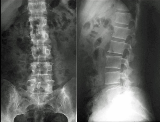
A 47-year-old female entering menopause presented to the outpatient Orthopaedic clinic of our hospital (June, 2001) with a 2-year history (July, 1999) of constant low back pain radiating to her buttocks. The pain was exaggerated when walking or standing but was slightly better when lying in bed. The pain disturbed her during sleep from time to time. A local health care provider had initially diagnosed (April, 2000) mild osteoarthritic changes at the L2–L3 level of the lumbar spine. He reported that on physical examination, there was neither any notable accompanying symptom, such as local tenderness, pattern of radiation, and neurovascular deficit, nor any abnormal findings in the urine test analysis. A potential pathological lumbar fracture, secondary to osteoporosis, was diagnosed 5 months ago after a new private consultation. She additionally complained of lower limb weakness. Plain radiographs by that time (January, 2001), including the chest, lumbar spine, and pelvis, were diagnosed with no abnormal findings (Figs. 1 and 2).

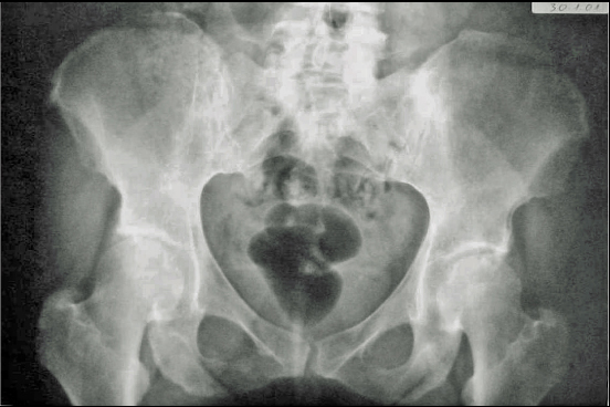
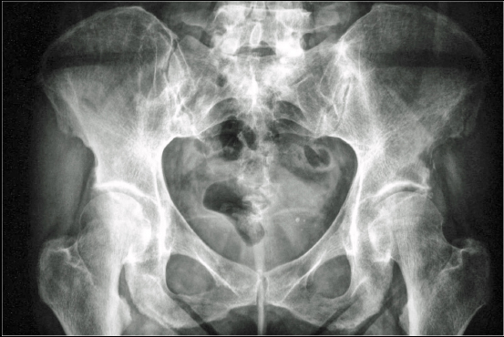
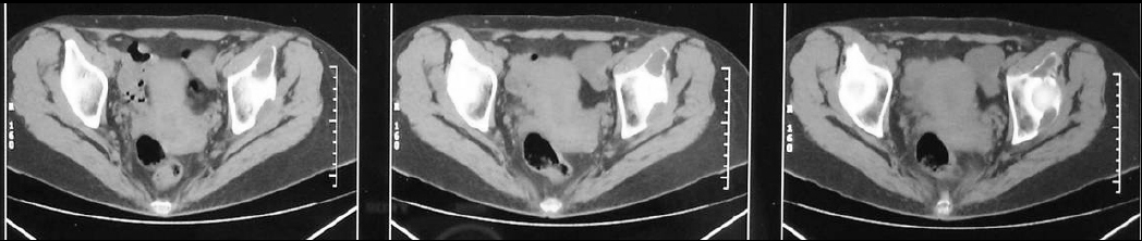
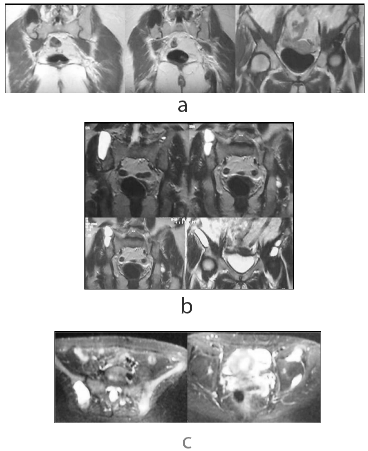
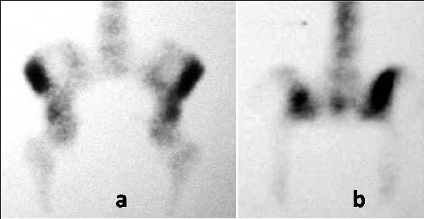
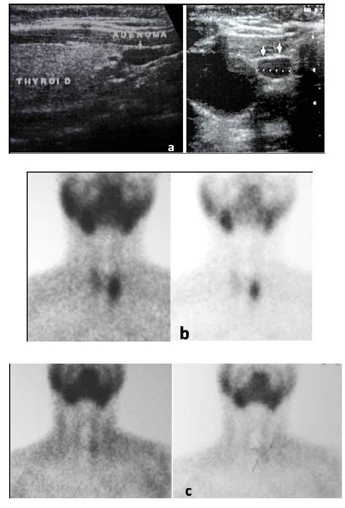
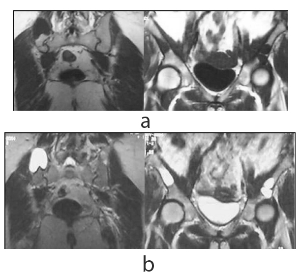
On presentation, the physical examination revealed left hip pain, more pronounced in the last degrees of internal rotation, and right sacroiliac pain. Specific areas of tenderness were localized below the left anterior iliac crest and laterally of the right sacroiliac joint. A new radiograph of the pelvis indicated a lytic lesion above the left acetabulum and another bigger one that was surrounded by a sclerotic rim laterally to the right sacroiliac joint (Fig. 3). A pelvic CT scan revealed that the two well-defined osteolytic lesions affected both the cortical and trabecular microstructure. They were both relatively well demarcated and exhibited bone erosion and cortical attenuation with no evidence of expansion or invading soft tissue (Fig. 4). A subsequent pelvic MRI showed that the lesions were hypointense on T1-weighted images, hyperintense on T2-weighted images, and exhibited the fluid-fluid level appearance, potentially due to intratumoral haemorrhage. The findings were considered consistent with those of an aneurysmal bone cyst, but the appearance of two lesions indicated the potential diagnosis of skeletal or systemic (presenting with typical skeletal and splenic involvement) cystic angiomatosis (Fig. 5). A 99mTc-MDP whole-body bone scan was performed (July, 2001), and it demonstrated elevated uptake in both lesions (Fig. 6). Laboratory studies were then performed (Table 1). The diagnosis of pHPT was established on the basis of the biochemical findings. Imaging studies, including ultrasound and 99mTc sestamibi scans, of the parathyroid glands followed (January, 2002), and they revealed a left inferior parathyroid adenoma (Fig. 7a-c). The patient had a standard parathyroid operation 3 months later (April, 2002), the tumor was removed, and histopathology revealed a parathyroid adenoma. The patient was admitted 4 days postsurgery to the Department of Internal Medicine of our hospital with the diagnosis of postoperative hypoparathyroidism and HBS. The clinical symptoms and signs of hypocalcaemia included numbness and tingling (paraesthesia) in the perioral area as well as in the hands and feet, muscle cramps, and carpopedal spasms. The Trousseau sign of latent tetany, in which the wrist and metacarpophalangeal joints flex, the proximal and distal interphalangeal joints extend, and the fingers adduct, was positive. The sign is also known as ‘main d’accoucheur’ (French for ‘hand of the obstetrician’) because it supposedly resembles the position of an obstetrician’s hand in delivering a baby. There was a paleness of the skin and conjunctivae. Abnormal vaginal bleeding was also diagnosed. Laboratory findings revealed persistent hypocalcaemia. Treatment included blood transfusion, uterine curettage, intravenous calcium infusion, as well as oral administration of calcitriol, calcium supplement, and magnesium. The patient recovered and was discharged from the hospital after 40 days. New pelvic MRI 3 years later (November, 2004) revealed no change in the appearance of the brown tumors (Fig. 8a, b). During the final follow-up (2021), the biochemical parameters, including serum calcium, vitamin D, and PTH, were normal but the pelvic radiograph indicated no significant changes in the appearance of the brown tumors (Fig. 9).

Table 1. Laboratory examination data of the patient.
Laboratory Studies 20/07/2001 02/10/2001 11/01/2002
Haematocrit 29.4% (36-46) - 29% (36-46)
Haemoglobin 9.9 gr% (12-14) - 9.5 gr% (12-14)
RBC 3.2 M/μl (4,2-5,4) - 3.6 M/μl (4,2-5,4)
ESR 35 mm/1st hour - 61 mm/1st hour
Total proteins 8.7 gr% (6-8) - -
Albumin 5.6 gr% (4-5.5) - -
Globulin 3.1 gr% (2-3) - -
ALP 922 U/L (100-290) 1185 IU/L (80-300) 1332 U/L (100-290)
Calcium Total 1.94 mgr% (0.98-1.27) Ionized 2.9 meg/l (2.1-2.7)
Total 12.5 mg/dl (8.3-10.4)
Total 1.93 mgr% (0.98-1.27)
24-hour urine calcium - - 289.5 mg/24h (25-250)
PTH - - 1042 pg/ml (10-71)
WBC, platelets, Blood sugar, Serum urea, creatinine, phosphate, immunoglobulins, C3, C4, anti-nuclear, anti-DNA, anti-smooth muscle, anti-mitochondrial antibodies, Urine test Normal values - -
RBC: Red Blood Count; ESR: Erythrocyte Sedimentation Rate; ALP: Alkaline Phosphatase; PTH: Parathormone; WBC: White Blood Count; C: Complement.
Fig. (1). Lumbar spine radiographs indicated no abnormal findings.

Fig. (2). Anteroposterior pelvic radiograph was diagnosed with no abnormal findings.

Fig. (3). Anteroposterior pelvic radiograph indicating two poorly defined osteolytic lesions in the left supra-acetabular region and adjacent to the right sacroiliac joint.

Fig. (4). CT scanning showed that the left supra-acetabular lesion was associated with structural reabsorption of the iliac wing.

Fig. (5). MRI showed two low signal intensity pelvic lesions on T1-weighted post-contrast (intravenous gadolinium) images (a). The tumors showed high signal intensity on T2-weighted (b) and short tau inversion recovery images (c).

Fig. (6). Whole-body bone scintigraphy with 99mTc-MDP, showing an irregular ring of increased uptake of the tracer in both pelvic lesions, on anterior (a) and posterior (b) views.

Fig. (7). Parathyroid ultrasound (a) showed a homogeneously hypoechoic nodule overlying the thyroid gland, while 99mTc sestamibi scans 10 minutes after radiotracer injection (early phase) (b), and 3 hours post-administration of the radioactive tracer (c), demonstrated pathological intense uptake in a definite area localized in the left inferior parathyroid gland.

Fig. (8). MRI using T1- (a) and T2-weighted (b) images revealed no regression of the pelvic brown tumors 3 years after surgical removal of the parathyroid adenoma.

Fig. (9). The radiographic appearance of the pelvic brown tumors has not changed significantly after a 20-year follow-up.

CONCLUSION

A high index of clinical suspicion may be required for the diagnosis of pHPT in a patient presenting in the common orthopaedic practice. A minimal increase of the serum calcium, a mild increase of the ESR, and a significant increase of total ALP were the only initial laboratory findings in the reported case, which had already developed brown tumors of the pelvis.

CONSENT FOR PUBLICATION

Permission to use data and images has been obtained from the patient.

FUNDING

The author received no financial support for this study.

CONFLICT OF INTEREST

The author certifies that he has no commercial associations (such as consultancies, stock ownership, equity interest, patent/licensing arrangements, etc.) that might pose a conflict of interest in connection with the submitted article.

ACKNOWLEDGEMENTS

Declared none.

REFERENCES

[1] Petti GH Jr. Hyperparathyroidism. Otolaryngol Clin North Am 1990; 23(2): 339-55.
[2] Ahmad R, Hammond JM. Primary, secondary, and tertiary hyperparathyroidism. Otolaryngol Clin North Am 2004; 37(4): 701-13. vii-viii
[3] Fraser WD. Hyperparathyroidism. Lancet 2009; 374(9684): 145-58.
[4] Cordellat IM. Hyperparathyroidism: Primary or secondary disease? Reumatol Clin 2012; 8(5): 287-91.
[5] Yuzawa Y, Watanabe Y. Hypercalcemia is the most common manifestation of hyperparathyroidism. Jpn J Clin Med 1995; 53(4): 864-9.
[6] Silverberg SJ, Bilezikian JP. Primary hyperparathyroidism: still evolving? J Bone Miner Res 1997; 12(5): 856-62.
[7] al Zahrani A, Levine MA. Primary hyperparathyroidism. Lancet 1997; 349(9060): 1233-8.
[8] Miedlich S, Krohn K, Paschke R. Update on genetic and clinical aspects of primary hyperparathyroidism. Clin Endocrinol (Oxf) 2003; 59(5): 539-54.
[9] Silverberg SJ, Bilezikian JP. Asymptomatic primary hyperparathyroidism: A medical perspective. Surg Clin North Am 2004; 84(3): 787-801.
[10] Mack LA, Pasieka JL. Asymptomatic primary hyperparathyroidism: A surgical perspective. Surg Clin North Am 2004; 84(3): 803-16.
[11] Bilezikian JP, Brandi ML, Rubin M, Silverberg SJ. Primary hyperparathyroidism: New concepts in clinical, densitometric and biochemical features. J Intern Med 2005; 257(1): 6-17.
[12] Colognesi A, de Tullio D, Messina F, Ferrocci G, Stano R, Azzena G. Primary hyperparathyroidism related to a parathyroid adenoma: The dramatic clinical evolution of a misdiagnosed patient and its surgical solution. Minerva Chir 2006; 61(1): 51-6.
[13] Habib Z, Camacho P. Primary hyperparathyroidism: An update. Curr Opin Endocrinol Diabetes Obes 2010; 17(6): 554-60.
[14] Rubin MR, Manavalan JS, Dempster DW, et al. Parathyroid hormone stimulates circulating osteogenic cells in hypoparathyroidism. J Clin Endocrinol Metab 2011; 96(1): 176-86.
[15] Amaral LM, Queiroz DC, Marques TF, Mendes M, Bandeira F. Normocalcemic versus hypercalcemic primary hyperparathyroidism: More stone than bone? J Osteoporos 2012; 2012128352
[16] Khan AA, Hanley DA, Rizzoli R, et al. Primary hyperparathyroidism: review and recommendations on evaluation, diagnosis, and management. A Canadian and international consensus. Osteoporos Int 2017; 28(1): 1-19.
[17] Yavropoulou MP, Poulios C, Michalopoulos N, et al. A role for circular non-coding RNAs in the pathogenesis of sporadic parathyroid adenomas and the impact of gender-specific epigenetic regulation. Cells 2018; 8(1): 15.
[18] Blanchard C, Mirallié E, Mathonnet M. Sporadic primary hyperparathyroidism. J Visc Surg 2010; 147(5): e285-95.
[19] Walker MD, Bilezikian JP. Primary hyperparathyroidism: recent advances. Curr Opin Rheumatol 2018; 30(4): 427-39.
[20] Mantzoros I, Kyriakidou D, Galanos-Demiris K, et al. A rare case of primary hyperparathyroidism caused by a giant solitary parathyroid adenoma. Am J Case Rep 2018; 19: 1334-7.
[21] Insogna KL. Primary Hyperparathyroidism. N Engl J Med 2018; 379(11): 1050-9.
[22] Choy KW. Primary Hyperparathyroidism. N Engl J Med 2018; 379(25)e43
[23] Bilezikian JP, Bandeira L, Khan A, Cusano NE. Hyperparathyroidism. Lancet 2018; 391(10116): 168-78.
[24] Yavropoulou MP, Anastasilakis AD, Panagiotakou A, Kassi E, Makras P. Gender predilection in sporadic parathyroid adenomas. Int J Mol Sci 2020; 21(8): 2964.
[25] Karpati G, Frame B. Neuropsychiatric disorders in primary hyperparathyroidism: Clinical analysis with review of the literature. Arch Neurol 1964; 10: 387-97.
[26] Kistler H. Primary hyperparathyroidism. An analysis of 152 patients with special references to acute life threatening complications (acute hyperparathyroidism). Schweiz Med Wochenschr 1976; 106(Suppl. 3): 1-61.
[27] Palmer FJ. The clinical manifestations of primary hyperparathyroidism. Compr Ther 1983; 9(2): 56-64.
[28] Bardin T. Primary hyperparathyroidism. Clinical, epidemiological and histological aspects. Ann Endocrinol (Paris) 1994; 55(5): 143-6.
[29] Sugimoto T. Osteoporosis associated with primary hyperparathyroidism. Jpn J Clin Med 2003; 61(2): 299-304.
[30] Walker MD, Rubin M, Silverberg SJ. Nontraditional manifestations of primary hyperparathyroidism. J Clin Densitom 2013; 16(1): 40-7.
[31] Gasser RW. Clinical aspects of primary hyperparathyroidism: Clinical manifestations, diagnosis, and therapy. Wien Med Wochenschr 2013; 163(17-18): 397-402.
[32] Verdelli C, Corbetta S. Mechanisms in endocrinology: Kidney involvement in patients with primary hyperparathyroidism: An update on clinical and molecular aspects. Eur J Endocrinol 2017; 176(1): R39-52.
[33] Walker MD, Silverberg SJ. Primary hyperparathyroidism. Nat Rev Endocrinol 2018; 14(2): 115-25.
[34] Chiodini I, Cairoli E, Palmieri S, Pepe J, Walker MD. Non classical complications of primary hyperparathyroidism. Best Pract Res Clin Endocrinol Metab 2018; 32(6): 805-20.
[35] Imanishi Y. Secondary osteoporosis. Secondary osteoporosis by primary hyperparathyroidism. Clin Calcium 2018; 28(12): 1627-34.
[36] Anastasilakis DA, Makras P, Polyzos SA, Anastasilakis AD. Asymptomatic and normocalcemic hyperparathyroidism, the silent attack: A combo-endocrinology overview. Hormones (Athens) 2019; 18(1): 65-70.
[37] Mollerup CL, Vestergaard P, Frøkjaer VG, Mosekilde L, Christiansen P, Blichert-Toft M. Risk of renal stone events in primary hyperparathyroidism before and after parathyroid surgery: controlled retrospective follow up study. BMJ 2002; 325(7368): 807.
[38] Corbetta S, Baccarelli A, Aroldi A, et al. Risk factors associated to kidney stones in primary hyperparathyroidism. J Endocrinol Invest 2005; 28(2): 122-8.
[39] Sorokin I, Mamoulakis C, Miyazawa K, Rodgers A, Talati J, Lotan Y. Epidemiology of stone disease across the world. World J Urol 2017; 35(9): 1301-20.
[40] Cong X, Shen L, Gu X. Current opinions on nephrolithiasis associated with primary hyperparathyroidism. Urolithiasis 2018; 46(5): 453-7.
[41] Lemos ALP, Andrade SRL, Pontes LLH, et al. High rate of occult urolithiasis in normocalcemic primary hyperparathyroidism. Kidney Blood Press Res 2019; 44(5): 1189-95.
[42] Castellano E, Attanasio R, Boriano A, Borretta G. Clinical presentation of primary hyperparathyroidism in older adults. J Endocr Soc 2019; 3(12): 2305-12.
[43] Lowe H, McMahon DJ, Rubin MR, Bilezikian JP, Silverberg SJ. Normocalcemic primary hyperparathyroidism: Further characterization of a new clinical phenotype. J Clin Endocrinol Metab 2007; 92(8): 3001-5.
[44] Quesada Gómez JM. Diagnostic evaluation and differential diagnosis of primary hyperparathyroidism. Endocrinol Nutr 2009; 56(Suppl. 1): 14-9.
[45] Adami S, Marcocci C, Gatti D. Epidemiology of primary hyperparathyroidism in Europe. J Bone Miner Res 2002; 17(Suppl. 2): N18-23.
[46] Silverberg SJ. Vitamin D deficiency and primary hyperparathyroidism. J Bone Miner Res 2007; 22(Suppl. 2): V100-4.
[47] Mosekilde L. Primary hyperparathyroidism and the skeleton. Clin Endocrinol (Oxf) 2008; 69(1): 1-19.
[48] Lindeman BM, Pesce CE, Tsai HL, et al. Lower vitamin D levels in surgical hyperparathyroidism versus thyroid patients. Am Surg 2014; 80(5): 505-10.
[49] Walker MD, Cong E, Lee JA, et al. Low vitamin D levels have become less common in primary hyperparathyroidism. Osteoporos Int 2015; 26(12): 2837-43.
[50] Carsote M, Paduraru DN, Nica AE, Valea A. Parathyroidectomy: Is vitamin D a player for a good outcome? J Med Life 2016; 9(4): 348-52.
[51] Villiaumey J, Hioco D, Chanzy MO, Chigot JP, Mellière D, Proye C. The new clinical picture of primary hyperparathyroidism. Diagnostic circumstances and current symptomatic characteristics. Results of a multicenter study. Rev Rhum Mal Osteoartic 1988; 55(8): 561-8.
[52] Heath H III. Clinical spectrum of primary hyperparathyroidism: evolution with changes in medical practice and technology. J Bone Miner Res 1991; 6(Suppl. 2): S63-70.
[53] Silverberg SJ. Natural history of primary hyperparathyroidism. Endocrinol Metab Clin North Am 2000; 29(3): 451-64.
[54] Bilezikian JP. Primary hyperparathyroidism. Endotext Feingold KR, Anawalt B, Boyce A, et al. 2017.
[55] Baleva R, Todorov T. Primary hyperparathyroidism in childhood and adolescence. Vutr Boles 1982; 21(2): 137-42.
[56] Kollars J, Zarroug AE, van Heerden J, et al. Primary hyperparathyroidism in pediatric patients. Pediatrics 2005; 115(4): 974-80.
[57] Steiniche T, Christiansen P, Vesterby A, et al. Primary hyperparathyroidism: bone structure, balance, and remodeling before and 3 years after surgical treatment. Bone 2000; 26(5): 535-43.
[58] Christiansen P. The skeleton in primary hyperparathyroidism: A review focusing on bone remodeling, structure, mass, and fracture. APMIS Acta Pathol Microbiol Immunol Scand, Suppl 2001; 102(102): 1-52.
[59] Montagnani A, Gonnelli S, Cepollaro C, et al. Graphic trace analysis of ultrasound at the phalanges may differentiate between subjects with primary hyperparathyroidism and with osteoporosis: A pilot study. Osteoporos Int 2002; 13(3): 222-7.
[60] De Geronimo S, Romagnoli E, Diacinti D, D’Erasmo E, Minisola S. The risk of fractures in postmenopausal women with primary hyperparathyroidism. Eur J Endocrinol 2006; 155(3): 415-20.
[61] Vignali E, Viccica G, Diacinti D, et al. Morphometric vertebral fractures in postmenopausal women with primary hyperparathyroidism. J Clin Endocrinol Metab 2009; 94(7): 2306-12.
[62] Lewiecki EM. Management of skeletal health in patients with asymptomatic primary hyperparathyroidism. J Clin Densitom 2010; 13(4): 324-34.
[63] Marcocci C, Cianferotti L, Cetani F. Bone disease in primary hyperparathyrodism. Ther Adv Musculoskelet Dis 2012; 4(5): 357-68.
[64] Stein EM, Silva BC, Boutroy S, et al. Primary hyperparathyroidism is associated with abnormal cortical and trabecular microstructure and reduced bone stiffness in postmenopausal women. J Bone Miner Res 2013; 28(5): 1029-40.
[65] Imai H, Katagiri M. Bone mineral quantifying of methods in hyperparathyroidism. Jpn J Clin Med 1995; 53(4): 885-9.
[66] Uchiyama T, Tanizawa T, Ito A, Endo N, Takahashi HE. Microstructure of the trabecula and cortex of iliac bone in primary hyperparathyroidism patients determined using histomorphometry and node-strut analysis. J Bone Miner Metab 1999; 17(4): 283-8.
[67] Khosla S, Melton J III. Fracture risk in primary hyperparathyroidism. J Bone Miner Res 2002; 17(Suppl. 2): N103-7.
[68] Lewiecki EM, Miller PD. Skeletal effects of primary hyperparathyroidism: bone mineral density and fracture risk. J Clin Densitom 2013; 16(1): 28-32.
[69] Ben Hassine L, Lahmar L, Bouaziz M, et al. Vertebral collapse revealing primary hyperparathyroidism in a 14-year-old girl. Arch Pediatr 2016; 23(4): 373-7.
[70] Liu M, Williams J, Kuo J, Lee JA, Silverberg SJ, Walker MD. Risk factors for vertebral fracture in primary hyperparathyroidism. Endocrine 2019; 66(3): 682-90.
[71] Nordenström E, Westerdahl J, Lindergård B, Lindblom P, Bergenfelz A. Multifactorial risk profile for bone fractures in primary hyperparathyroidism. World J Surg 2002; 26(12): 1463-7.
[72] Eriksen EF. Primary hyperparathyroidism: Lessons from bone histomorphometry. J Bone Miner Res 2002; 17(Suppl. 2): N95-7.
[73] Vu TD, Wang XF, Wang Q, et al. New insights into the effects of primary hyperparathyroidism on the cortical and trabecular compartments of bone. Bone 2013; 55(1): 57-63.
[74] Chalmers J, Irvine GB. Fractures of the femoral neck in elderly patients with hyperparathyroidism. Clin Orthop Relat Res 1988; (229): 125-30.
[75] Lafforgue P, Pham T, Denizot A, Daumen-Legré V, Acquaviva PC. Bone insufficiency fractures as an inaugural manifestation of primary hyperparathyroidism. Rev Rhum Engl Ed 1996; 63(7-8): 475-9.
[76] Deshmukh RG, Alsagoff SA, Krishnan S, Dhillon KS, Khir AS. Primary hyperparathyroidism presenting with pathological fracture. J R Coll Surg Edinb 1998; 43(6): 424-7.
[77] Morgan G, Ganapathi M, Afzal S, Grant AJ. Pathological fractures in primary hyperparathyroidism. A case report highlighting diagnostic difficulties. Injury 2002; 33(3): 288-91.
[78] Wu PH, Wang CJ. Normocalcemic primary hyperparathyroidism with fractures. J Arthroplasty 2002; 17(6): 805-9.
[79] Verlaan L, van der Wal B, de Maat GJ, Walenkamp G, Nollen-Lopez L, van Ooij A. Primary hyperparathyroidism and pathological fractures: A review. Acta Orthop Belg 2007; 73(3): 300-5.
[80] Souza ER, Scrignoli JA, Bezerra FC, Ribeiro SL, Passos LF. Devastating skeletal effects of delayed diagnosis of complicated primary hyperparathyroidism because of ectopic adenoma. J Clin Rheumatol 2008; 14(5): 281-4.
[81] Ostojić Z, Grle M, Moro G, Zubak Z, Ostojić M. Bone fractures in a 53-year-old patient with parathyroid adenoma: A case report. Coll Antropol 2010; 34(Suppl. 1): 295-8.
[82] Cormier C. Fracture risk in hyperparathyroidism. Joint Bone Spine 2012; 79(3): 216-8.
[83] Dutta P, Mandal A, Bandyopadhyay U, Santra S, Sarkar PS. Management of pathological fracture in patients with primary hyperparathyroidism. J Indian Med Assoc 2013; 111(12): 835-6.
[84] Olatoke SA, Agodirin OS, Rahman GA. Serial pathologic fractures of five long bones on four separate occasions in a patient with primary hyperparathyroidism, challenges of management in a developing country: A case report. Pan Afr Med J 2013; 15: 45.
[85] French R, Oweis D, Kurup V. Bilateral femoral fractures in a 21-year-old man following a simple fall. BMJ Case Rep 2013; 2013
[86] Bandeira F, Cusano NE, Silva BC, et al. Bone disease in primary hyperparathyroidism. Arq Bras Endocrinol Metabol 2014; 58(5): 553-61.
[87] Alattas MH, Dimentberg R. Multiple fractures in a 22-year-old man after a simple fall. J Surg Case Rep 2015; 2015(10)rjv117
[88] Benameur Y, Guerrouj H, Ghfir I, Ben Rais Aouad N. Unusual pathological fracture of the clavicle revealing primary hyperparathyroidism: A case report. J Med Case Reports 2017; 11(1): 342.
[89] Merali F, Leung M. Bilateral pathologic mid-tibial stress fractures: Initial presentation of mild primary hyperparathyroidism. Can Fam Physician 2017; 63(8): 610-2.
[90] Rider T, Baig I, Sayer C, Crown A. Pathological finger fracture. BMJ 2013; 346: f1024.
[91] Ozaki A, Tanimoto T, Yamagishi E. Finger fractures as an early manifestation of primary hyperparathyroidism among young patients: A case report of a 30-year-old male with recurrent osteoporotic fractures. Medicine (Baltimore) 2016; 95(20)e3683
[92] Lancourt JE, Hochberg F. Delayed fracture healing in primary hyperparathyroidism. Clin Orthop Relat Res 1977; (124): 214-8.
[93] Sauvé PS, Suliman IG, Calder JD. Primary hyperparathyroidism presenting as delayed fracture union. Knee Surg Sports Traumatol Arthrosc 2009; 17(5): 551-4.
[94] Kinoshita M, Naito K, Aritomi K. Forearm nonunion caused by hyperparathyroidism with 7 years follow up: A case report. Int J Surg Case Rep 2017; 38: 158-62.
[95] Searfoss R, Tripi J, Bowers W. Triceps brachii rupture: case report. J Trauma 1976; 16(3): 244-6.
[96] Golinval A, Ledoux JP, Jodogne G, Nizet M, Meurisse M, Lejeune G. Pathological fractures and tendon ruptures connected with primary and secondary hyperparathyroidism. Apropos of 7 cases. Acta Orthop Belg 1982; 48(6): 885-97.
[97] Lavalle C, Aparicio LA, Moreno J, Chavez de los Rios J, Robles-Paramo A, Fraga A. Bilateral avulsion of quadriceps tendons in primary hyperparathyroidism. J Rheumatol 1985; 12(3): 596-8.
[98] Chen CH, Niu CC, Yang WE, Chen WJ, Shih CH. Spontaneous bilateral patellar tendon rupture in primary hyperparathyroidism. Orthopedics 1999; 22(12): 1177-9.
[99] Tsourvakas S, Gouvalas K, Gimtsas C, Tsianas N, Founta P, Ameridis N. Bilateral and simultaneous rupture of the triceps tendons in chronic renal failure and secondary hyperparathyroidism. Arch Orthop Trauma Surg 2004; 124(4): 278-80.
[100] Salah MM, Yong YR, Poh WT, Chong LR. Multiple spontaneous tendon ruptures from enthesis failure in primary hyperparathyroidism: A case report and review of imaging findings. Skeletal Radiol 2019; 48(8): 1279-87.
[101] Sturdee SW, Bollen SR. Intra-articular calcification in primary hyperparathyroidism. Knee 2004; 11(4): 323-5.
[102] Bone LB, Roach JW, Ward WT, Worthen HG. Slipped capital femoral epiphysis associated with hyperparathyroidism. J Pediatr Orthop 1985; 5(5): 589-92.
[103] Kinoshita J, Kaneda K, Matsuno T, Hosokawa Y, Nagashio R. Slipped capital femoral epiphysis associated with hyperparathyroidism. A case report. Int Orthop 1995; 19(4): 245-7.
[104] Yang WE, Shih CH, Wang KC, Jeng LB. Slipped capital femoral epiphyses in a patient with primary hyperparathyroidism. J Formos Med Assoc 1997; 96(7): 549-52.
[105] El Scheich T, Marquard J, Westhoff B, et al. Approach to the management of slipped capital femoral epiphysis and primary hyperparathyroidism. J Pediatr Endocrinol Metab 2012; 25(5-6): 407-12.
[106] Kumar G, Mathew V, Kandathil JC, Theruvil B. Primary hyperparathyroidism presenting as slipped capital femoral epiphysis. Postgrad Med J 2020; 96(1134): 235-6.
[107] Kaiser W. Osseous course of primary hyperparathyroidism. Beitr Orthop Traumatol 1970; 17(2): 148-50.
[108] Sundaram M, Scholz C. Primary hyperparathyroidism presenting with acute paraplegia. AJR Am J Roentgenol 1977; 128(4): 674-6.
[109] Kaplan FS, Soffer SR, Fallon MD, Haddad JG, Dalinka M, Raffensperger EC. Osteomalacia as a very late manifestation of primary hyperparathyroidism. Clin Orthop Relat Res 1988; (228): 26-32.
[110] Silverberg SJ, Shane E, de la Cruz L, et al. Skeletal disease in primary hyperparathyroidism. J Bone Miner Res 1989; 4(3): 283-91.
[111] Parisien M, Silverberg SJ, Shane E, Dempster DW, Bilezikian JP. Bone disease in primary hyperparathyroidism. Endocrinol Metab Clin North Am 1990; 19(1): 19-34.
[112] Daras M, Georgakopoulos T, Avdelidis D, Gravani A, Tuchman AJ. Spinal cord compression in primary hyperparathyroidism. Report of a case and review of the literature. Spine 1990; 15(3): 238-40.
[113] Bassler T, Wong ET, Brynes RK. Osteitis fibrosa cystica simulating metastatic tumor. An almost-forgotten relationship. Am J Clin Pathol 1993; 100(6): 697-700.
[114] Khan A, Bilezikian J. Primary hyperparathyroidism: pathophysiology and impact on bone. CMAJ 2000; 163(2): 184-7.
[115] Syed Z, Khan A. Skeletal effects of primary hyperparathyroidism. Endocr Pract 2000; 6(5): 385-8.
[116] Hsieh MC, Ko JY, Eng HL. Pathologic fracture of the distal femur in osteitis fibrosa cystica simulating metastatic disease. Arch Orthop Trauma Surg 2004; 124(7): 498-501.
[117] Mustonen AO, Kiuru MJ, Stahls A, Bohling T, Kivioja A, Koskinen SK. Radicular lower extremity pain as the first symptom of primary hyperparathyroidism. Skeletal Radiol 2004; 33(8): 467-72.
[118] Yang Q, Sun P, Li J, et al. Skeletal lesions in primary hyperparathyroidism. Am J Med Sci 2015; 349(4): 321-7.
[119] Misiorowski W, Czajka-Oraniec I, Kochman M, Zgliczyński W, Bilezikian JP. Osteitis fibrosa cystica-a forgotten radiological feature of primary hyperparathyroidism. Endocrine 2017; 58(2): 380-5.
[120] Makras P, Anastasilakis AD. Bone disease in primary hyperparathyroidism. Metabolism 2018; 80: 57-65.
[121] Meindok H, Rapoport A, Oreopoulos DG, Rabinovich S, Meema HE, Meema S. Quantitative radionuclide scanning in metabolic bone disease. Nucl Med Commun 1985; 6(3): 141-8.
[122] Joyce JM, Idea RJ, Grossman SJ, Liss RG, Lyons JB. Multiple brown tumors in unsuspected primary hyperparathyroidism mimicking metastatic disease on radiograph and bone scan. Clin Nucl Med 1994; 19(7): 630-5.
[123] Ishikawa S, Ozaki T, Kawai A, Inoue H, Doihara H. Hyperparathyroid crisis in a patient with a giant brown tumor of the iliac bone: A case report. Hiroshima J Med Sci 1998; 47(1): 27-30.
[124] Kalambokis G, Economou G, Kamina S, Papachristou DJ, Bai M, Tsianos EV. Multiple brown tumors of the ribs simulating malignancy. J Endocrinol Invest 2005; 28(8): 738-40.
[125] Triantafillidou K, Zouloumis L, Karakinaris G, Kalimeras E, Iordanidis F. Brown tumors of the jaws associated with primary or secondary hyperparathyroidism. A clinical study and review of the literature. Am J Otolaryngol 2006; 27(4): 281-6.
[126] Diamanti-Kandarakis E, Livadas S, Tseleni-Balafouta S, et al. Brown tumor of the fibula: unusual presentation of an uncommon manifestation. Report of a case and review of the literature. Endocrine 2007; 32(3): 345-9.
[127] Khalil PN, Heining SM, Huss R, et al. Natural history and surgical treatment of brown tumor lesions at various sites in refractory primary hyperparathyroidism. Eur J Med Res 2007; 12(5): 222-30.
[128] Jouan A, Zabraniecki L, Vincent V, Poix E, Fournié B. An unusual presentation of primary hyperparathyroidism: severe hypercalcemia and multiple brown tumors. Joint Bone Spine 2008; 75(2): 209-11.
[129] Hoshi M, Takami M, Kajikawa M, et al. A case of multiple skeletal lesions of brown tumors, mimicking carcinoma metastases. Arch Orthop Trauma Surg 2008; 128(2): 149-54.
[130] Demay MB, Rosenthal DI, Deshpande V. Case records of the Massachusetts General Hospital. Case 16-2008. A 46-year-old woman with bone pain. N Engl J Med 2008; 358(21): 2266-74.
[131] Proimos E, Chimona TS, Tamiolakis D, Tzanakakis MG, Papadakis CE. Brown tumor of the maxillary sinus in a patient with primary hyperparathyroidism: A case report. J Med Case Reports 2009; 3: 7495.
[132] Su AW, Chen CF, Huang CK, Chen PC, Chen WM, Chen TH. Primary hyperparathyroidism with brown tumor mimicking metastatic bone malignancy. J Chin Med Assoc 2010; 73(3): 177-80.
[133] Ruggeri RM, Calamoneri E, Russo A, et al. Supra-acetabular brown tumor due to primary hyperparathyroidism associated with chronic renal failure. ScientificWorldJournal 2010; 10: 799-805.
[134] Ergen FB, Ayvaz M, Yildiz AE, Aydingoz U, Gedikoglu G. Brown tumour presenting as a soft-tissue mass. Clin Radiol 2012; 67(3): 286-9.
[135] Ullah E, Ahmad M, Ali SA, Redhu N. Primary hyperparathyroidism having multiple Brown tumors mimicking malignancy. Indian J Endocrinol Metab 2012; 16(6): 1040-2.
[136] Yu HI, Lu CH. Sacroiliitis-like pain as the initial presentation of primary hyperparathyroidism. Arch Osteoporos 2012; 7: 315-8.
[137] Das KJ, Sehgal AK, Jaiman A, Sethi RS. Osteosclerotic and osteolytic manifestations of hyperparathyroidism in a case of Tc99m SestaMIBI positive parathyroid adenoma. Indian J Nucl Med 2015; 30(3): 263-5.
[138] Panagopoulos A, Tatani I, Kourea HP, Kokkalis ZT, Panagopoulos K, Megas P. Osteolytic lesions (brown tumors) of primary hyperparathyroidism misdiagnosed as multifocal giant cell tumor of the distal ulna and radius: A case report. J Med Case Reports 2018; 12(1): 176.
[139] Park YJ, Yoon TR, Park KS, Ko JW. Subchondral bone restoration of supra-acetabular brown tumor secondary to parathyroid carcinoma: A case report. Hip Pelvis 2018; 30(2): 120-4.
[140] Hu J, He S, Yang J, Ye C, Yang X, Xiao J. Management of brown tumor of spine with primary hyperparathyroidism: A case report and literature review. Medicine (Baltimore) 2019; 98(14)e15007
[141] Hamidi S, Mottard S, Berthiaume MJ, Doyon J, Bégin MJ, Bondaz L. Brown tumor of the iliac crest initially misdiagnosed as a giant cell tumor of the bone. Endocrinol Diabetes Metab Case Rep 2020; 2020: 20-0029.
[142] Bennett J, Suliburk JW, Morón FE. Osseous manifestations of primary hyperparathyroidism: Imaging findings. Int J Endocrinol 2020; 20203146535
[143] Davies AM, Evans N, Mangham DC, Grimer RJ. MR imaging of brown tumour with fluid-fluid levels: A report of three cases. Eur Radiol 2001; 11(8): 1445-9.
[144] Takeshita T, Takeshita K, Abe S, Takami H, Imamura T, Furui S. Brown tumor with fluid-fluid levels in a patient with primary hyperparathyroidism: radiological findings. Radiat Med 2006; 24(9): 631-4.
[145] Hong WS, Sung MS, Chun KA, et al. Emphasis on the MR imaging findings of brown tumor: A report of five cases. Skeletal Radiol 2011; 40(2): 205-13.
[146] Xie C, Tsakok M, Taylor N, Partington K. Imaging of brown tumours: A pictorial review. Insights Imaging 2019; 10(1): 75.
[147] Chavda DV, Frock JT, Zielinski CM, Walla DJ, McGuire MH. Reversal of the histology of bone after parathyroidectomy in patients with hyperparathyroidism. J South Orthop Assoc 1998; 7(1): 65-71.
[148] Agarwal G, Mishra SK, Kar DK, et al. Recovery pattern of patients with osteitis fibrosa cystica in primary hyperparathyroidism after successful parathyroidectomy. Surgery 2002; 132(6): 1075-83.
[149] Fine EJ. Parathyroid imaging: its current status and future role. Semin Nucl Med 1987; 17(4): 350-9.
[150] McBiles M, Lambert AT, Cote MG, Kim SY. Sestamibi parathyroid imaging. Semin Nucl Med 1995; 25(3): 221-34.
[151] Nguyen BD. Parathyroid imaging with Tc-99m sestamibi planar and SPECT scintigraphy. Radiographics 1999; 19(3): 601-14.
[152] Smith JR, Oates ME. Radionuclide imaging of the parathyroid glands: patterns, pearls, and pitfalls. Radiographics 2004; 24(4): 1101-15.
[153] Palestro CJ, Tomas MB, Tronco GG. Radionuclide imaging of the parathyroid glands. Semin Nucl Med 2005; 35(4): 266-76.
[154] Cheung K, Wang TS, Farrokhyar F, Roman SA, Sosa JA. A meta-analysis of preoperative localization techniques for patients with primary hyperparathyroidism. Ann Surg Oncol 2012; 19(2): 577-83.
[155] Moralidis E. Radionuclide parathyroid imaging: A concise, updated review. Hell J Nucl Med 2013; 16(2): 125-33.
[156] Romanidis K, Karathanos E, Nagorni EA, et al. Parathyroid adenoma detected with 99mTc-tetrofosmin dual-phase scintigraphy: A case report. BMC Res Notes 2014; 7: 335.
[157] Garas G, Poulasouchidou M, Dimoulas A, Hytiroglou P, Kita M, Zacharakis E. Radiological considerations and surgical planning in the treatment of giant parathyroid adenomas. Ann R Coll Surg Engl 2015; 97(4): e64-6.
[158] Butt HZ, Husainy MA, Bolia A, London NJ. Ultrasonography alone can reliably locate parathyroid tumours and facilitates minimally invasive parathyroidectomy. Ann R Coll Surg Engl 2015; 97(6): 420-4.
[159] Xue J, Liu Y, Ji T, et al. Comparison between technetium-99m methoxyisobutylisonitrile scintigraphy and ultrasound in the diagnosis of parathyroid adenoma and parathyroid hyperplasia. Nucl Med Commun 2018; 39(12): 1129-37.
[160] Vu TH, Schellingerhout D, Guha-Thakurta N, et al. Solitary parathyroid adenoma localization in technetium Tc99m sestamibi SPECT and multiphase multidetector 4D CT. AJNR Am J Neuroradiol 2019; 40(1): 142-9.
[161] Kowa XY, Richards P, Waterhouse M, Parvanta L, Adams A. Atypical presentations of parathyroid gland pathology: A pictorial review. Eur J Radiol Open 2019; 6: 320-9.
[162] Mazokopakis EE, Skarakis SI. Investigating patients with normocalcemic hyperparathyroidism: When is a parathyroid scintigraphy indicated? World J Nucl Med 2019; 18(3): 227-31.
[163] Wong W, Foo FJ, Lau MI, Sarin A, Kiruparan P. Simplified minimally invasive parathyroidectomy: A series of 100 cases and review of the literature. Ann R Coll Surg Engl 2011; 93(4): 290-3.
[164] Sahsamanis G, Gkouzis K, Samaras S, Pinialidis D, Dimitrakopoulos G. Surgical management of a giant parathyroid adenoma through minimal invasive parathyroidectomy. A case report. Int J Surg Case Rep 2017; 31: 262-5.
[165] Mizamtsidi M, Nastos C, Mastorakos G, et al. Diagnosis, management, histology and genetics of sporadic primary hyperparathyroidism: old knowledge with new tricks. Endocr Connect 2018; 7(2): R56-68.
[166] Silverberg SJ, Clarke BL, Peacock M, et al. Current issues in the presentation of asymptomatic primary hyperparathyroidism: proceedings of the Fourth International Workshop. J Clin Endocrinol Metab 2014; 99(10): 3580-94.
[167] Singhal S, Johnson CA, Udelsman R. Primary hyperparathyroidism: what every orthopedic surgeon should know. Orthopedics 2001; 24(10): 1003-9.
[168] Adámek S, Libánský P, Lischke R, Foltán R, Kubinyi J, Broulík P. Surgical therapy of primary hyperparathyrodism in the context of orthopaedic diagnosis and treatment: our experiences in 441 patients. Acta Chir Orthop Traumatol Cech 2011; 78(4): 355-60.
[169] Brasier AR, Nussbaum SR. Hungry bone syndrome: clinical and biochemical predictors of its occurrence after parathyroid surgery. Am J Med 1988; 84(4): 654-60.
[170] Farese S. The hungry bone syndrome-an update. Ther Umsch 2007; 64(5): 277-80.
[171] Witteveen JE, van Thiel S, Romijn JA, Hamdy NA. Hungry bone syndrome: still a challenge in the post-operative management of primary hyperparathyroidism: A systematic review of the literature. Eur J Endocrinol 2013; 168(3): R45-53.
[172] Ghilardi G, De Pasquale L. Hungry bone syndrome after parathyroidectomy for primary hyperthyroidism. Surgery Curr Res 2014; 4: 168.
[173] Jain N, Reilly RF. Hungry bone syndrome. Curr Opin Nephrol Hypertens 2017; 26(4): 250-5.
[174] Florakis D, Karakozis S, Tseleni-Balafouta S, Makras P. Lessons learned from the management of hungry bone syndrome following the removal of an atypical parathyroid adenoma. J Musculoskelet Neuronal Interact 2019; 19(3): 379-84.
[175] Ko WC, Liu CL, Lee JJ, Liu TP, Wu CJ, Cheng SP. Osteocalcin is an independent predictor for hungry bone syndrome after parathyroidectomy. World J Surg 2020; 44(3): 795-802.
[176] Guillén Martínez AJ, Smilg Nicolás C, Moraleda Deleito J, Guillén Martínez S, García-Purriños García F. Risk factors and evolution of calcium and parathyroid hormone levels in hungry bone syndrome after parthyroidectomy for primary hyperparathyroidism. Endocrinol Diabetes Nutr 2020; 67(5): 310-6.
[177] Cartwright C, Anastasopoulou C. Hungry bone syndrome. StatPearls 2020.