EDITORIAL


Clinical and Radiographic Features of Phalangeal Osteoid Osteoma


N. K. Sferopoulos1, *
1 2nd Department of Orthopaedic Surgery, Aristotle University of Thessaloniki, ‘G. Gennimatas’ Hospital, 546 35, Thessaloniki, Greece


Article Metrics

CrossRef Citations:
0
Total Statistics:

Full-Text HTML Views: 1481
Abstract HTML Views: 298
PDF Downloads: 279
ePub Downloads: 203
Total Views/Downloads: 2261
Unique Statistics:

Full-Text HTML Views: 836
Abstract HTML Views: 203
PDF Downloads: 221
ePub Downloads: 156
Total Views/Downloads: 1416


Creative Commons License
© 2021 N. K. Sferopoulos

open-access license: This is an open access article distributed under the terms of the Creative Commons Attribution 4.0 International Public License (CC-BY 4.0), a copy of which is available at: (https://creativecommons.org/licenses/by/4.0/legalcode). This license permits unrestricted use, distribution, and reproduction in any medium, provided the original author and source are credited.

* Address correspondence to this author at the 2nd Department of Orthopaedic Surgery, Aristotle University of Thessaloniki, “G. Gennimatas” Hospital, 546 35, Thessaloniki, Greece; Tel: 00302310963270; Fax: 00302310968265; E-mail: sferopoulos@yahoo.com


EDITORIAL

The purpose of this editorial is to perform an extensive review of the relevant international literature about the clinical and radiographic features of phalangeal osteoid osteomas of the hand or foot, to indicate the value of clinical suspicion for the early diagnosis since they commonly appear with atypical findings, and to present four illustrative cases, two adults and two children.

Osteoid osteoma is a benign osteoblastic tumor of unknown origin. It usually affects children, adolescents and young adults showing a strong male predominance. The tumor has a predilection for the metaphysis or diaphysis of long bones of the lower limbs but it may occur anywhere in the skeleton. It typically consists of a well-demarcated radiolucent rarefied area of bone-forming and prostaglandin-secreting nidus, occupying the midcortex, which is surrounded by an extensive fusiform reactive sclerotic host bone. Reactive sclerosis is usually more pronounced in children. The lesion may also be found in the medulla (spongiosa) of bone. In the cortical presentation, it may rarely occupy the endosteal or subperiosteal surface of the cortex. Intramedullary tumors are usually surrounded by a thin rim of reactive sclerosis and may frequently be periarticular or intraarticular, presenting with synovitis and joint effusion [1-10].

Approximately 10% of osteoid osteomas involve the hand. Of them, 6% occur in the phalanges, 2% in the metacarpals and 2% in the carpal bones. Cortical localization of the tumor is usually seen in the phalanges and metacarpals, while intramedullary lesions are usually encountered in the carpal bones [11-23]. In the fingers, the proximal phalanx is the most typically affected site, while lesions of the distal phalanx are rarely encountered. The index finger is the most commonly involved digit, while the thumb is the least affected one [24-41]. Phalangeal lesions of the foot account for about 2% of all cases [42-47].

In an accessible bone, swelling over an osteoid osteoma is a typical finding. In a phalangeal osteoid osteoma, swelling may be localized or generalized, resembling dactylitis or macrodactyly. Whenever the lesion affects the terminal phalanx, enlargement of the entire fingertip, producing clubbing, appears to be very likely [24, 41, 48-54]. The presentation of clubbing in a digit should raise the suspicion of a bone tumor, even when plain radiographs show only soft tissue swelling or vague radiographic changes [55]. Both the enlargement of the finger and the hypertrophy of the nail may be explained by the increased focal vascularity [49]. Osteoid osteomas localized in the subungual area have also been reported [56]. An accompanying erythema of the overlying skin may rarely be detected. Cases associated with either erythema or nail deformities may easily mislead the diagnosis [57-59].

Nocturnal pain and exquisite tenderness over the site of the nidus are the most constant clinical features of phalangeal osteoid osteomas of the hand or foot. Pain is unrelated to use or motion and it may precede the radiographic changes by many months [60]. The severity of pain may increase gradually, so that it may take months or even more than a year before a patient seeks medical advice. The pain is typically worse at night and is usually relieved by small doses of acetylsalicylic acid (aspirin) or nonsteroidal anti-inflammatory drugs (NSAIDs). The use of both may be gradually less effective in reducing pain to an acceptable level. On occasions, pain may not be worse at night or may not be relieved at all by these medications. The appearance of an atypical type of pain (dull, vague or diffuse) and the absolute lack of pain are exceptionally rare in non-phalangeal osteoid osteomas. Painless osteoid osteomas appear in the digits more frequently than in any other skeletal location [56, 61-64]. Vasomotor disturbances, namely increased skin temperature in the affected region, and profuse digital tip perspiration or intense autonomic phenomena, which may mimic peripheral nerve compression or reflex sympathetic dystrophy have also been reported [48].

A common diagnostic problem of juxtaarticular or intraarticular phalangeal osteoid osteomas is that they may present as monoarticular interphalangeal arthritis with decreased range of motion (ROM). This presentation may also misdirect the diagnosis of a phalangeal osteoid osteoma to monoarticular juvenile idiopathic arthritis [55, 65-68].

Conventional radiographs remain the most important imaging study of the suspected lesion. Plain radiograps may demonstrate the typical finding of a phalangeal osteoid osteoma, which is a radiolucent nidus, usually less than 1 cm at its greatest dimension, localized within the cortex. Nidus identification is of decisive importance for diagnosis and treatment. Surrounding dense sclerosis or extensive periosteal reaction may overshadow the nidus, indicating the diagnosis of an obscure phalangeal osteoid osteoma [55, 69-71]. Such cases may be confused with malignancy [72]. One theory to account for the discrepancy in findings between studies is the possibility that the nidus simply becomes more visible with time [37, 55]. With time, the nidus matures and becomes calcified and more radiopaque. Ossification of the nidus usually starts from the center, producing a central zone of density surrounded by a ringlike band of radiolucent zone (ring sequestrum sign or annular sequestrum of Jaffe) [2, 73, 74]. The radiographic sign of sclerosis may take many years after excision of the nidus to subside, but eventually, it does [75]. Unlike osteoid osteomas of the long bones that show clear demarcation between the nidus and the reactive bone, phalangeal lesions may not be well defined [24, 33, 50, 55]. A relative absence of dense reactive bone surrounding the nidus is a common radiographic appearance of a cortical phalangeal lesion [37, 50, 51, 55, 76]. Cases with relative absence of sclerosis are more likely to indicate a diagnosis of subacute osteomyelitis [77]. Local osteopenia from disuse and the intraarticular location of the lesion have been proposed as potential causes for the lack of reactive sclerosis [33, 76]. Periosteal reaction may be evident in 65% of cases. The lack of periosteal reaction may be due to the close proximity of the tumor to the interphalangeal joint [37], while in lesions localized to the distal phalanges may be due to the poorly developed periosteum in the region [55]. The presence of thickened Sharpey’s fibres may also explain the absence of periosteal reaction associated with osteoid osteomas in the distal phalanges [59, 78]. Phalangeal osteoid osteoma with a double-nidus has also been reported [79].

The diagnosis of intramedullary, subperiosteal or periarticular [73, 80-90] phalangeal osteoid osteomas is particularly difficult since both the clinical symptoms and the radiographic findings may be minimal, absent or unspecific. Intramedullary osteoid osteomas are usually well-circumscribed, the reactive bone surrounding the nidus is less pronounced and the nidus may be partially or completely calcified. Subperiosteal or exostotic osteoid osteoma is the rarest type and usually presents as a soft tissue mass adjacent to or eroding the bony cortex. Reactive sclerosis is much more pronounced than in intramedullary and periarticular lesions [30, 81, 83].

Conventional tomography may serve as a valued adjunct to plain radiography. A 3-phase Technetium-99m MDP bone scan is of considerable value since it demonstrates the intense focal increase in uptake by the nidus. Once the lesion has been localized by bone scan, cross sectional and three-dimensional imaging with Computed Tomography (CT) is usually used. Magnetic Resonance Imaging (MRI) will demonstrate the soft tissue and bone marrow edema but is rarely needed, because CT both better demonstrates the nidus and aids in differentiating between the nidus and reactive bone. The use of angiography may also result in improved recognition of the nidus [75, 91-94]. Atypical histological features may also be often encountered in phalangeal osteoid osteomas in both children and adults, such as ill demarcation between the nidus and reactive bone, and absence of dense reactive bone surrounding the nidus [55, 95-102].

The main differential diagnosis in a patient with a painful fingertip swelling may include whitlow or paronychia, osteomyelitis, glomus tumour, subungual exostosis, osteochondroma, enchondroma, osteoid osteoma, and arthritis [29, 50, 55, 56, 59], In addition, in cases with a painful nontraumatic swelling of the proximal or middle phalanx differential diagnosis includes soft tissue lesions, bone abscess and osteomyelitis of acute or chronic sclerosing types, monoarticular rheumatoid arthritis, enostosis (bone island), stress fracture, benign bone tumors, and malignancy [23, 30, 78, 79, 103, 104].

There has been no report of malignant transformation of this neoplasm so far. A complication of phalangeal osteoid osteoma, which has rarely been reported, is premature physeal fusion [49, 105, 106]. In a two children report, in the one of them local damage to the phalangeal physeal plate by the lesion could be excluded [107]. Therefore, other etiological factors were considered, such as the local increase in vascularity or the regional osteoporosis [108], in a similar way that they were encountered in epiphyses around the knee in treated tuberculosis of the hip [109]. Distant recurrence of the tumor is extremely rare with only one reported case [110]. Local recurrence of the clinical symptoms and a new nidus will form following inadequate treatment [91, 111, 112]. Cases localized in a juxtacortical area of the articular surface that necessitate removal of an articular area are more prone to complications [75].

Four patients with a histologically proven phalangeal osteoid osteoma of a finger or toe are presented. The clinical findings were assessed for localized or generalized swelling, for the presence/type of pain and for interphalangeal arthritis associated with decreased ROM. The evaluated radiographic features were identification of a radiolucent nidus, internal matrix within the nidus, sclerosis surrounding the nidus, periosteal reaction, periosteal/cortical sclerosis, and cortical erosion.

There were two adult male patients, 21 (patient 1) and 23 (patient 2) years of age, respectively in Figs. (1 and 2). Two children, a 12-year-old boy (patient 3) and a 13-year-old girl (patient 4) were also identified (Figs. 3 and 4). In the two adult patients, the tumor involved the thumb and middle finger, respectively. In the two children, the great toe and the index finger were involved, respectively. The tumor was localized in the distal portion of the proximal phalanx in all cases. Swelling, pain and local tenderness were the presenting symptoms. Swelling was localized in 3 patients and generalized, in the form of dactylitis, only in patient 1. The cardinal feature of night pain as well as local tenderness over the nidus was evident in all cases. However, complete relief of pain with the use of aspirin or other NSAIDs was noted only in patient 1. No erythema or local increase in temperature was detected in any of the patients. The adjacent proximal interphalangeal joint was very tender and had limited ROM in all cases. Precise localization of the nidus was determined in patients 1, 2 and 3 with conventional radiography and in patient 4 with MRI. An intracortical localization of the nidus was noted in patients 2 and 3, while an intramedullary one was noted in patients 1 and 4. A distinctive area of sclerosis surrounding the lytic lesion was seen only in the adult patients. Minimal and absent sclerosis were evident in patients 3 and 4, respectively. Internal sclerosing matrix within the nidus and the ring sequestrum sign were evident on the radiographs in the adult patients, while in patients 3 and 4, both findings were delineated with the use of CT and MRI, respectively. Cortical/periosteal sclerosis was present in patients 1, 2 and 4. Cortical erosion as well as local osteopenia was not identified in any of the patients.

Fig. (1). A 21-year-old man with a 12-month history of painful swelling of the left thumb. Anteroposterior and lateral radiographs showed an intramedullary lytic lesion in the distal portion of the proximal phalanx of the thumb. Internal sclerosing matrix was evident within the lesion. A broad zone of reactive bone surrounded the lesion. Periosteal as well as cortical sclerosis of the proximal phalanx of the thumb was also noted.

Fig. (2). A 23-year-old man with an 18-month history of a painful swelling in the proximal phalanx of the right middle finger. Anteroposterior and lateral radiographs indicated a lytic lesion in the distal part of the radial cortex of the proximal phalanx of the middle finger. Its center was almost completely occupied by sclerosing matrix. A broad zone of reactive bone surrounded the lesion. Periosteal as well as cortical sclerosis of the distal, radial cortex of the proximal phalanx of the middle finger was also evident.

Fig. (3). A 12-year-old boy with a 6-month history of a painful swelling of the proximal phalanx of the left great toe. Clinical appearance of the patient’s feet, showing enlargement of the proximal phalanx of the left great toe. At radiography, a lytic lesion surrounded by mild sclerosis was evident on the distal portion of the proximal phalanx of the great toe. CT indicated the presence of an intracortical lytic lesion with internal sclerosing matrix.

Fig. (4). A 13-year-old girl with a 2-month history of painful swelling of the proximal phalanx of the left index finger. Anteroposterior and lateral radiographs revealed periosteal reaction of the proximal phalanx of the index finger but no other radiographic evidence of a bone lesion. Fat suppressed MRI view indicating a lytic lesion with internal sclerosing matrix on the distal portion of the proximal phalanx. Inflammatory soft tissue changes as well as synovitis and effusion of the proximal interphalangeal joint were evident.

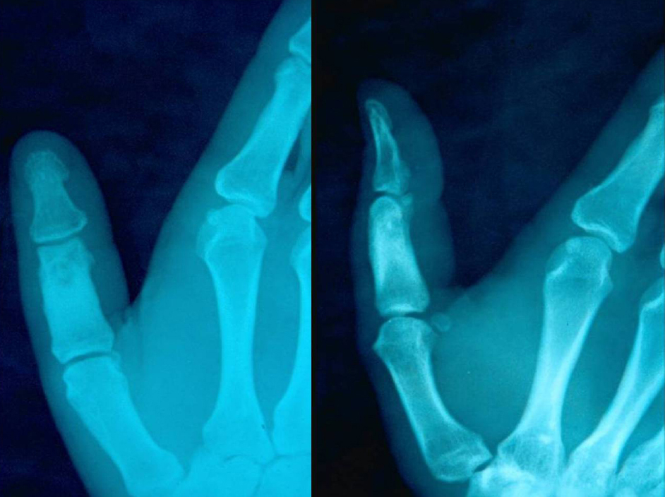
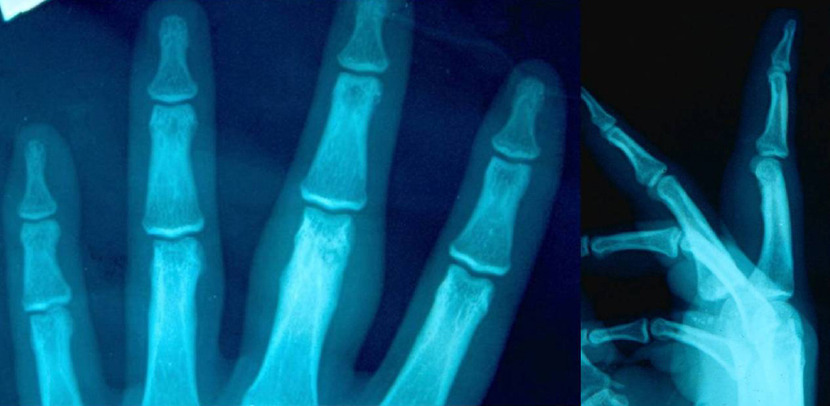
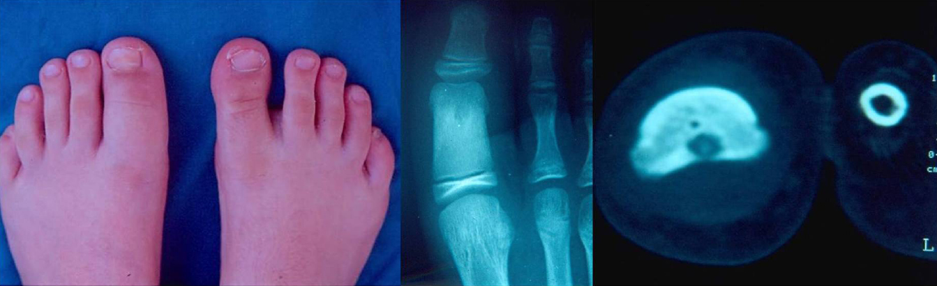
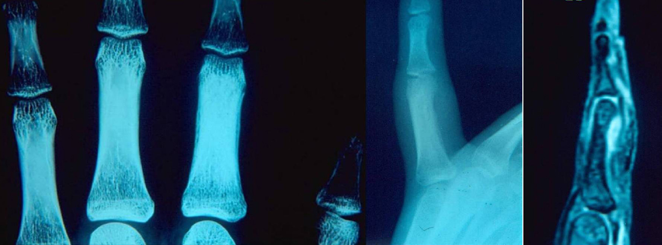
The clinical and the radiographic findings in both adult patients indicated the diagnosis of a phalangeal osteoid osteoma. However, the clinical and radiographic evaluation in both children yielded nonspecific results. Thus, the diagnosis of osteomyelitis could not be excluded in patient 3, while in patient 4 the findings were not sufficient to make a differential diagnosis from osteomyelitis or malignancy.

CONCLUSION

It has strongly been emphasized in the literature that phalangeal osteoid osteoma may commonly present with atypical clinical and/or radiographic findings in both adults and children. In such cases, clinical suspicion remains the most sensitive indicator for diagnosis. Furthermore, imaging evaluation including conventional tomography, bone scintigraphy, CT, which has the highest specificity, and MRI is mandatory in patients presenting with atypical or no radiographic findings as well as in patients with atypical clinical findings, such as a phalangeal painless swelling, clubbing of the fingertip, dactylitis or macrodactyly, and monoarticular interphalangeal arthritis. Awareness of the potential atypical clinical and/or radiographic characteristics of the phalangeal osteoid osteoma of the hand or foot may lead to earlier diagnosis and treatment in both adults and children.

FUNDING

None.

CONFLICT OF INTEREST

The author declares no conflict of interest, financial or otherwise.

ACKNOWLEDGEMENTS

Declared none.

REFERENCES

[1] Freiberger RH, Loitman BS, Helpern M, Thompson TC. Osteoid osteoma; a report on 80 cases. Am J Roentgenol Radium Ther Nucl Med 1959; 82(2): 194-205.
[2] Jackson RP, Reckling FW, Mants FA. Osteoid osteoma and osteoblastoma. Similar histologic lesions with different natural histories. Clin Orthop Relat Res 1977; (128): 303-13.
[3] Swee RG, McLeod RA, Beabout JW. Osteoid osteoma. Detection, diagnosis, and localization. Radiology 1979; 130(1): 117-23.
[4] Smith FW, Gilday DL. Scintigraphic appearances of osteoid osteoma. Radiology 1980; 137(1 Pt 1): 191-5.
[5] Rand JA, Sim FH, Unni KK. Two osteoid-osteomas in one patient. A case report. J Bone Joint Surg Am 1982; 64(8): 1243.
[6] Mullin DM, Rodan BA, Bean WJ, Sehayik S, Sehayik R. Osteoid osteoma in unusual locations: detection and diagnosis. South Med J 1986; 79(10): 1299-301.
[7] Kransdorf MJ, Stull MA, Gilkey FW, Moser RP Jr. Osteoid osteoma. Radiographics 1991; 11(4): 671-96.
[8] Lee EH, Shafi M, Hui JHP. Osteoid osteoma: a current review. J Pediatr Orthop 2006; 26(5): 695-700.
[9] Malik AA, Trevalyan S, Khan WS, Hughes A, Hill JC, Redfern DR. New techniques for localisation and excision of osteoid osteoma. J Hand Surg Eur Vol 2008; 33(3): 389-91.
[10] Czinner M, Kebrle R, Matějovský Z, Němejc M. Osteoid osteoma of the upper extremity. Acta Chir Orthop Traumatol Cech 2019; 86(1): 77-82.
[11] Testa G, Mazzoleni G. Osteoid osteoma; case with phalangeal localization. Arch Radiol 1951-1952-1952; 27(4): 417-26.
[12] Carroll RE. Osteoid osteoma in the hand. J Bone Joint Surg Am 1953; 35(4): 888-93.
[13] Dunitz NL, Lipscomb PR, Ivins JC. Osteoid osteoma of the hand and wrist. Am J Surg 1957; 94(1): 65-9.
[14] Aube L, Roy L, Lessard R. Osteoid osteoma of the phalanges. Can Med Assoc J 1968; 98(9): 455-8.
[15] Sullivan M. Osteoid osteoma of the fingers. Hand 1971; 3(2): 175-8.
[16] Braun S, Chevrot A, Tomeno B, et al. Phalangeal osteoid osteoma (13 cases). Rev Rhum Mal Osteoartic 1979; 46(4): 225-33.
[17] McConnell B III, Dell PC. Localization of an osteoid osteoma nidus in a finger by use of computed tomography: a case report. J Hand Surg Am 1984; 9A(1): 139-41.
[18] Doyle LK, Ruby LK, Nalebuff EG, Belsky MR. Osteoid osteoma of the hand. J Hand Surg Am 1985; 10(3): 408-10.
[19] Ambrosia JM, Wold LE, Amadio PC. Osteoid osteoma of the hand and wrist. J Hand Surg Am 1987; 12(5 Pt 1): 794-800.
[20] Kozlowski K, Azouz EM, Campbell J, et al. Primary bone tumours of the hand. Report of 21 cases. Pediatr Radiol 1988; 18(2): 140-8.
[21] De Smet L, Fabry G. Osteoid Osteoma of the hand and carpus: Peculiar presentations and imaging. Acta Orthop Belg 1995; 61(2): 113-6.
[22] Bilgin SS, Yildiz Y, Güçlü B, Sağlik Y. Osteoid osteoma in the hand: an evaluation of eight patients. Acta Orthop Traumatol Turc 2004; 38(3): 206-11.
[23] Hamdi MF, Tarhouni L, Daghfous M, Bergaoui N, Baccari S. Osteoid osteoma of the phalanx and metacarpal bone: report of 17 cases. Musculoskelet Surg 2015; 99(1): 61-5.
[24] Giannikas A, Papachristou G, Tiniakos G, Chrysafidis G, Hartofilakidis-Garofalidis G. Osteoid osteoma of the terminal phalanges. Hand 1977; 9(3): 295-300.
[25] Grundberg AB. Osteoid osteoma of the thumb: report of a case. J Hand Surg Am 1977; 2(4): 266.
[26] Levy Y, Rosenheck S, Greiff M, Torok G. Osteoid osteoma of the distal phalanx of the thumb. Acta Orthop Scand 1979; 50(6 Pt 1): 667-9.
[27] Ramesh P, Khan F, Kamboj H. Painful lesions at the fingertips. J R Soc Med 2004; 97(1): 30-1.
[28] Sproule JA, Khan F, Fogarty EE. Osteoid osteoma: painful enlargement of the second toe. Arch Orthop Trauma Surg 2004; 124(5): 354-6.
[29] Di Gennaro GL, Lampasi M, Bosco A, Donzelli O. Osteoid osteoma of the distal thumb phalanx: a case report. Chir Organi Mov 2008; 92(3): 179-82.
[30] Tsang DS, Wu DY. Osteoid osteoma of phalangeal bone. J Formos Med Assoc 2008; 107(7): 582-6.
[31] Prietzel T, Hitzler P, Wojan M, Aigner T, von Salis-Soglio G. Painful enlargement of the 2nd toe due to an osteoid osteoma in the distal phalanx. Z Orthop Unfall 2009; 147(3): 362-5.
[32] Ebrahimzadeh MH, Omidi-Kashani F, Hoseini MR. Painful and tender toe, osteoid osteoma of the distal phalanx of toe, a diagnostic dilemma. Foot 2009; 19(4): 232-5.
[33] Galdi B, Capo JT, Nourbakhsh A, Patterson F. Osteoid osteoma of the thumb: A case report. Hand (N Y) 2010; 5(4): 423-6.
[34] Hattori H, Takase K, Morohashi A. Osteoid osteoma of the great toe. Orthopedics 2011; 34(8): e432-5.
[35] Rotzer A, Umbricht R, von Wartburg U. Post-traumatic osteoid osteoma of the hand. A rare cause of chronic pain. Case report and review of the literature. Handchir Mikrochir Plast Chir 1998; 30(5): 335-7.
[36] Andalib A, Sajadie-Khajouei S. Osteoid osteoma of distal phalanx: A rare disorder and review of literature. J Res Med Sci 2013; 18(3): 264-6.
[37] Kotnis N, James SL. Imaging features of osteoid osteoma of the phalanges. Skeletal Radiol 2015; 44(10): 1461-6.
[38] Feron M, Desdoits A, Bronfen C, Jeanne-Pasquier C, Haumont T. Osteoid osteoma on distal phalanx tip of second toe, without scintigraphy fixation. Arch Pediatr 2015; 22(12): 1279-83.
[39] Durgia B, Jain A, Agarwal S. Osteoid osteoma of distal phalanx of middle finger-A diagnostic dilemma. J Hand Surg Asian Pac Vol 2016; 21(3): 395-8.
[40] Horiuchi K, Horiuchi Y, Ochi K. Osteoid osteoma of the distal phalanx of the ring finger with clubbed finger deformity: A case report. J Hand Surg Asian Pac Vol 2017; 22(2): 248-50.
[41] Bailey JR, Holbrook J. Phalangeal osteoid osteoma of thumb. J Hand Surg Am 2019; 44(11): 995.e1-4.
[42] Kahn MD, Tiano FJ, Lillie RC. Osteoid osteoma of the great toe. J Foot Surg 1983; 22(4): 325-8.
[43] Nechvolodova OL, Zatsepin ST, Melkonian VM. Osteoid osteoma of the hands and feet. Ortop Travmatol Protez 1989; 1(1): 13-5.
[44] Onoue K, Kudawara I. Osteoid osteoma with cartilage formation of the distal phalanx in the toe. Orthopedics 2007; 30(8): 670-1.
[45] Jowett CR, Singh D. Osteoid osteoma of the great toe: A case report. Foot Ankle Surg 2010; 16(2): e12-5.
[46] Xarchas KC, Kyriakopoulos G, Manthas S, Oikonomou L. Hallux osteoid osteoma: A case report and literature review. Open Orthop J 2017; 11: 1066-72.
[47] Kurmis AP. Hallux osteoid osteoma: A case report and literature review. Open Orthop J 2019; 13: 22-3.
[48] Nakatsuchi Y, Sugimoto Y, Nakano M. Osteoid osteoma of the terminal phalanx. J Hand Surg [Br] 1984; 9(2): 201-3.
[49] Rosborough D. Osteoid osteoma. Report of a lesion in the terminal phalanx of a finger. J Bone Joint Surg Br 1966; 48(3): 485-7.
[50] Foucher G, Lemarechal P, Citron N, Merle M. Osteoid osteoma of the distal phalanx: a report of four cases and review of the literature. J Hand Surg [Br] 1987; 12(3): 382-6.
[51] Bowen CV, Dzus AK, Hardy DA. Osteoid osteomata of the distal phalanx. J Hand Surg [Br] 1987; 12(3): 387-90.
[52] McCarten GM, Dixon PL, Marshall DR. Osteoid osteoma of the distal phalanx: a case report. J Hand Surg [Br] 1987; 12(3): 391-3.
[53] Brown RE, Russell JB, Zook EG. Osteoid osteoma of the distal phalanx of the finger: a diagnostic challenge. Plast Reconstr Surg 1992; 90(6): 1016-21.
[54] Inagaki H, Inoue G. Osteoid osteoma of the distal phalanx. Orthopedics 1999; 22(11): 1093-4.
[55] Burger IM, McCarthy EF. Phalangeal osteoid osteomas in the hand: a diagnostic problem. Clin Orthop Relat Res 2004; (427): 198-203.
[56] Ekmekci P, Bostanci S, Erdoğan N, Akçaboy B, Gürgey E. A painless subungual osteoid osteoma. Dermatol Surg 2001; 27(8): 764-5.
[57] Lakkis S, Bazzi JS, Shabb NS. Osteoid osteoma of the proximal phalanx of a toe. A case report. Bull Hosp Jt Dis 1998; 57(2): 102-4.
[58] Barca F, Acciaro AL, Recchioni MD. Osteoid osteoma of the phalanx: enlargement of the toe--two case reports. Foot Ankle Int 1998; 19(6): 388-93.
[59] Becce F, Jovanovic B, Guillou L, Theumann N. Painful fingertip swelling of the middle finger. Osteoid osteoma of the distal phalanx of the middle finger. Skeletal Radiol 2011; 40(11): 1479-1480, 1501-1502.
[60] Fehring TK, Green NE. Negative radionuclide scan in osteoid osteoma. A case report. Clin Orthop Relat Res 1984; (185): 245-9.
[61] Lawrie TR, Aterman K, Sinclair AM. Painless osteoid osteoma. A report of two cases. J Bone Joint Surg Am 1970; 52(7): 1357-63.
[62] Wiss DA, Reid BS. Painless osteoid osteoma of the fingers-report of three cases. J Hand Surg Am 1983; 8(6): 914-7.
[63] Rex C, Jacobs L, Nur Z. Painless osteoid osteoma of the middle phalanx. J Hand Surg [Br] 1997; 22(6): 798-800.
[64] Themistocleous GS, Chloros GD, Benetos IS, Efstathopoulos DG, Gerostathopoulos NE, Soucacos PN. Osteoid osteoma of the upper extremity. A diagnostic challenge. Chir Main 2006; 25(2): 69-76.
[65] Kawaguchi Y, Sato C, Hasegawa T, Oka S, Kuwahara H, Norimatsu H. Intraarticular osteoid osteoma associated with synovitis: A possible role of cyclooxygenase-2 expression by osteoblasts in the nidus. Mod Pathol 2000; 13(10): 1086-91.
[66] Hedrich CM, Fiebig B, Sallmann S, et al. Osteoid osteomas of the fingers: An atypical localization? Two case reports and a review of the literature. Z Rheumatol 2008; 67(2): 145-148, 150.
[67] Rousié M, Sattari A, Schuind F. Mono-arthritis of the hand: Case report of osteoid osteoma. Rev Med Brux 2015; 36(1): 38-41.
[68] Vlaic J, Lamot L, Simunic S, Harjacek M, Bojic D. Unusual localization and presentation of osteoid osteoma mimicking juvenile spondyloarthritis: a case report. BMC Musculoskelet Disord 2019; 20(1): 17.
[69] Soler JM, Pizà G, Aliaga F. Special characteristics of osteoid osteoma in the proximal phalanx. J Hand Surg [Br] 1997; 22(6): 793-7.
[70] Johnson T, Steinbach L. Essentials of musculoskeletal imaging 2004.
[71] Bhusnurmath S, Hoch B. Benign bone-forming tumors.Current concepts in bone pathology Surgical pathology clinics 2012; 5(1): 101-6.
[72] Georgiev GP, Slavchev SA, Dimitrova IN. Osteoid osteoma of the middle phalanx of the third finger in a child mimicking a malignancy. Folia Med (Plovdiv) 2018; 60(2): 314-7.
[73] Muren C, Höglund M, Engkvist O, Juhlin L. Osteoid osteomas of the hand. Report of three cases and review of the literature. Acta Radiol 1991; 32(1): 62-6.
[74] Fechner RE, Mills SE. Tumors of the bones and joints.Atlas of tumor pathology 1993.
[75] Carroll R. Lesions of osseous origin.Tumors of the hand and upper limb 10th vol 1993.
[76] Kransdorf MJ, Murphey MD. Osseous tumours.Imaging of bone tumours and tumour-like lesions: Techniques and applications 2009.
[77] Meng QF, Watt I. Phalangeal osteoid osteoma. Br J Radiol 1989; 62(736): 321-5.
[78] Jones SN, Stoker DJ. Radiology at your fingertips; lesions of the terminal phalanx. Clin Radiol 1988; 39(5): 478-85.
[79] Allieu Y, Lussiez B, Benichou M, Cenac P. A double nidus osteoid osteoma in a finger. J Hand Surg Am 1989; 14(3): 538-41.
[80] Allieu Y, Lussiez B. L’osteome osteode au niveau de la main. Apropos de quarante-six cas. Osteoid osteoma in the hand: Report of a series of forty-six cases. Ann Chir Main 1988; 7: 298.
[81] Klein MJ, Parisien MV, Schneider-Stock R, Osteoid osteoma. In: Fletcher CDM, Unni KK, Mertens F, Eds. World Health organization classification of tumours.Pathology and genetics of tumours of soft tissue and bone 2002; 260-61.
[82] Amrami KK, Berger RA. Radiology corner: osteoid osteoma of the index finger: case presentation and discussion. J Hand Surg Am 2006; 31(2): 322-4.
[83] Kotnis NA, Davies AM, James SLJ. Hand and wrist.Imaging of bone tumours and tumour-like lesions: Techniques and applications 2009.
[84] Liu EH, Stone N, Alowami SO, Thoma A. Proximal phalanx osteoid osteoma: A case report and literature review. Plast Reconstr Surg Glob Open 2017; 5(5)e1332
[85] Noordin S, Allana S, Hilal K, et al. Osteoid osteoma: Contemporary management. Orthop Rev (Pavia) 2018; 10(3): 7496.
[86] Crosby LA, Murphy RP. Subperiosteal osteoid osteoma of the distal phalanx of the thumb. J Hand Surg Am 1988; 13(6): 923-5.
[87] Kayser F, Resnick D, Haghighi P, et al. Evidence of the subperiosteal origin of osteoid osteomas in tubular bones: Analysis by CT and MR imaging. AJR Am J Roentgenol 1998; 170(3): 609-14.
[88] LaCroix ML, Thomas JR, Nicholas RW. Subperiosteal osteoid osteoma of the distal phalanx of the fourth toe. Orthopedics 2001; 24(7): 695-6.
[89] Oztürk A, Yalçinkaya U, Ozkan Y, Yalçin N. Subperiosteal osteoid osteoma in the hallux of a 9-year-old female. J Foot Ankle Surg 2008; 47(6): 579-82.
[90] Shubinets V, Ben-Amotz O, Steinberg DR. A subperiosteal proximal phalanx osteoid osteoma: A challenging diagnosis. J Hand Surg Asian Pac Vol 2019; 24(2): 233-7.
[91] Huvos AG. Bone tumors: diagnosis, treatment and prognosis Atlas of orthopaedic pathology Philadelphia: WB Saunders 1979.
[92] Herring JA. Tachdjian’s pediatric orthopaedics 3rd ed. 2002.
[93] Shukla S, Clarke AW, Saifuddin A. Imaging features of foot osteoid osteoma. Skeletal Radiol 2010; 39(7): 683-9.
[94] French J, Epelman M, Johnson CM, Stinson Z, Meyers AB. MR imaging of osteoid osteoma: Pearls and pitfalls. Semin Ultrasound CT MR 2020; 41(5): 488-97.
[95] Resnick D. Tumors and tumorlike lesions of the bone.Diagnosis of bone and joint disorders 2nd ed. 1988.
[96] Mirra JM, Picci P, Gold RH. Bone tumors: Clinical, radiologic, and pathologic correlations 2nd ed. 1989.
[97] Unni KK. Dahlin’s bone tumours General aspects and data on 11087 cases 5th ed. 1997.
[98] Dorfman HD, Czerniak B. Bone Tumors 1998; 1261.
[99] Rosai J. Rosai and Ackerman’s surgical pathology 9th ed. 2004.
[100] Bullough PG. Orthopaedic pathology 4th ed. 2004.
[101] Resnick D, Kransdorf MJ. Tumour and tumour-like diseases. In: Resnick D, Ed. Bone and Joint Imaging 3rd ed. 2005.
[102] Heck RK. Benign bone tumors and nonneoplastic condition simulating bone tumors.Campbell’s operative orthopaedics 2008.
[103] Aghoutane EM, El Fezzazi R. [Osteoid osteoma of the phalanx in children and diagnostic problems: report of one case]. Chir Main 2012; 31(4): 199-201.
[104] Sferopoulos NK. Primary subacute hematogenous osteomyelitis in children. Orthopaedic Surgery and Traumatology 2017; 1(5): 148-57.
[105] Giustra PE, Freiberger RH. Severe growth disturbance with osteoid osteoma. A report of two cases involving the femoral neck. Radiology 1970; 96(2): 285-8.
[106] Bordelon RL, Cracco A, Book MK. Osteoid-osteoma producing premature fusion of the epiphysis of the distal phalanx of the big toe. A case report. J Bone Joint Surg Am 1975; 57(1): 120-2.
[107] Sevitt S, Horn JS. A painless and calcified osteoid osteoma of the little finger. J Pathol Bacteriol 1954; 67(2): 571-4.
[108] Spence AJ, Lloyd-Roberts GC. Regional osteoporosis in osteoid osteoma. J Bone Joint Surg Br 1961; 43(3): 501-7.
[109] Gill GG. The cause of discrepancy in length of the limbs following tuberculosis of the hip in children. J Bone Joint Surg 1944; 26: 272-81.
[110] Dahlin DC, Unni KK. Bone tumors General aspects and data on 8542 cases 4th ed. 1986.
[111] De Wet IS. Osteoid osteomata. Review of the literature with a report of five cases. S Afr J Surg 1967; 5(1): 13-24.
[112] Dunlop JA, Morton KS, Eliott GB. Recurrent osteoid osteoma. Report of a case with a review of the literature. J Bone Joint Surg Br 1970; 52(1): 128-33.