All published articles of this journal are available on ScienceDirect.

Incidence of Venous Thromboembolism in Patients Undergoing Major Hip Surgeries at a Single Institution: A Prospective Study
Abstract
Background:
Venous thoromboembolism (VTE) is one of the most significant complications after hip surgeries. Many studies have been reported about the incidence of VTE after THA, but a small number of reports were found concerning Periacetabular osteotomy, Revision THA and Surgery for hip fracture postoperatively. Furthermore, there exists no comparative study of the incidence of VTE among major hip surgeries at a single institution. We reported the incidence of VTE among hip surgeries performed at a single institution.
Methods:
A total of 820 Hip surgeries were performed at same institution. The procedures included 420 hips that underwent primary total hip arthroplasties (THA), 91 revision or removal of total hip arthroplasties (Revision THA), 144 periacetabular osteotomy (PAO) and 165 surgery for hip fracture (SHF) between 2006 and 2012. VTE was detected by Multidetector computed tomography (MDCT) that scanned 768 cases and by ultrasound that scanned 52cases postoperative 10-14 days.
Results:
The overall incidence of VTE was 12.2% (100 of 820). The incidence of VTE after THA was 13.1% (55 of 420), Revision THA was 13.2% (12 of 91), PAO was 2.1% (3 of 144) and SHF was 18.1% (30 of 165). The incidence of VTE was significantly higher in SHF than in PAO.
Conclusion:
This data indicates that the incidence of VTE after PAO is significantly lower than SHF and relatively lower than THA and Revision THA. A younger age and non-invasion of the bone marrow of the femur may have affected the result. Prophylaxis therapy was effective especially on SHF.
INTRODUCTION
Thromboembolism is a cause of morbidity and mortality for hospitalized patients [1]. Orthopedic surgeries of the hip joint such as total hip arthroplasties (THA), revision total hip arthroplasties (Revision THA), periacetabular osteotomy (PAO) and surgery for hip fracture (SHF) are commonly performed. However, patients undergoing major elective orthopedic surgery are in the highest risk category for thromboembolism (VTE). VTE includes deep venous thrombosis (DVT) and pulmonary embolism (PTE), is considered to be one of the most significant complications after primary THA, and a relatively large number of studies have been reported. However, a small number of reports have been found in the incidence of VTE concerning PAO, Revision THA and SHF postoperatively.
In an aging society, there will be a higher number of patients who receive THA. This indicates that there will be a higher number of patients who need revised THA potentially. This may evoke risk of complications postoperatively, including VTE. Developmental dysplasia of the hip with associated structural instability is a cause of secondary osteoarthritis. When this is left untreated, the hip degeneration will progress possibly to the point of end-stage disease. To prevent secondary osteoarthritis, various corrective hip osteotomy techniques have been described for the treatment of symptomatic dysplasia. We have performed modified periacetabular osteotomy without massive bone graft [2]. There exist a few reports concerning incidence of VTE after PAO. Hip fractures are common, especially in elder with osteoporosis. A patient with a hip fracture is exposed to a variety of risks and a significant mortality with or without operation. The complications include hip joint infection, pressure sores, myocardial infarction, urinary tract infection, pneumonia and VTE.
Most previous research has been limited to risk factors for VTE among patients undergoing THA. We focus on the incidence of VTE among major hip surgeries at the same institution, with almost the same protocol of rehabilitation, receiving routine thromboprophylaxis. To the best of our knowledge, there was no comparative study of VTE among four major hip surgeries performed at the same Hospital. The aim of this article is to investigate the incidence of VTE in patients operated THA, Revision THA, PAO and SHF at a single institution.
MATERIALS AND METHODS
We retrospectively studied patients who underwent hip surgeries (820 hips) at Asahikawa Medical University between 2006 and 2012. The study group included 168 men and 652 women. The procedures included 420 hips that underwent primary THA, 91 revised or removal of total hip arthroplasties (Revision THA), 144 PAO and 165 SHF at same institution (Table 1). 20 patients had multiple operations.
Sex, age, operation time and intraoperative blood loss of each operation.
| THA(420) | Rev.THA(91) | PAO(144) | SHF(165) | |
|---|---|---|---|---|
| Sex, male/female | 89/331 | 25/66 | 19/125 | 35/130 |
| Age, mean y±SD | 62.4±12.1 | 68.3±11.4 | 32.2±11.4 | 79.2±12.1 |
| Operation Time, mean min±SD | 61.4±20.5 | 126.9±51.3 | 99.7±25.4 | 55.0±27.3 |
| Intraoperative Blood loss, mean ml±SD | 296±220.9 | 647.9±522.1 | 449.9±320.8 | 115±229.2 |
Primary THA: all the procedures were performed through the posteolateral approach without trochanteric osteotomy. The mean age of the patients was 62.4±12.1 years. The mean time of operation was 61.4±20.5 min. The mean amount of intraoperative total blood loss was 296.3±220.9 ml. Revision THA: 72 hips had revision surgery and 19 hips were removed implant because of infection. 18 of 19 cases were referred to our hospital for definitive treatment. The mean age of the patients was 68.3±11.4 years. The mean time of operation was 126.9±51.3 min. The mean amount of intraoperative total blood loss was 647.9±522.1ml. PAO: 106 cases of Rotational acetabular osteotomy (RAO) and 38 cases of Chiari pelvic osteotomy (CPO) were performed through an Ollier lateral U approach along with a trochanteric osteotomy [3]. The greater trochanter with its tendinous insertion was then retracted proximally to expose the entire joint capsule. Then pelvic osteotomy was performed with use of reciprocating power saw (CPO) or with a curved osteotome (RAO). Two mm-diameter Kirschner wires were inserted from the proximal part of the ilium into the ischium to fix the fragment in position (CPO) or four bioabsorbable cortical screws were inserted to fix the acetabular fragment (RAO). The freed greater trochanter was reduced and fixed with two 6.5mm metallic cancellous screws. The mean age of the patients was 32.2±11.4 years. The mean time of operation was 99.7±25.4 min. The mean amount of intraoperative total blood loss was 449.9±320.8ml. SHF: surgical devices included 84 femoral short nail (FSN); 21 compression hip screws (CHS); 21 cannulated cancellous screws (CCHS) and 39 bipolar hip prostheses (BHP) with cement stem. The mean age of the patients was 79.2±12.1years. The mean time of operation was 55.0±27.3 min. The mean amount of intraoperative total blood loss was 115.5±229.2 ml.
All patients were treated with mechanical thromboprophylaxis (pressure stockings or compression devices) after the operation for three days. The patients who were not detected VTE preoperatively were applied compression devices and were not examined preoperatively applied pressure stockings because applying compression devices to the patients who have DVT preoperatively may course PTE. Active range of motion, quadriceps, and straight leg-raising exercise were encouraged immediately all the cases. All THA patients were allowed to be fully weight-bearing. For the patients who underwent PAO, partial weight-bearing was allowed postoperatively from 2006 to 2009. Since posterior column fractures were observed in several cases, non-weight-bearing walking was initially recommended for the first two weeks after surgery from 2010 to 2012 [4]. In most cases of Revision THA and SHF, weight-bearing as tolerated was allowed. In some cases weight-bearing avoided for several weeks and patients were encouraged to use a wheelchair postoperatively. No pharmacologic prophylaxis was used postoperatively from 2006 to 2009. Since 2010, patients who were not diagnosed as having renal or hepatic failure or a severe anemia received chemoprophylaxis therapy (Enoxaparin or Edoxaban) with mechanical thromboprophylaxis in this study. Total 306 of 820 patients received chemoprophylaxis therapy. 158 patients received chemoprophylaxis therapy with compression devices and 262 patients received compression device after THA. 19 patients received chemoprophylaxis therapy with compression devices and 72 patients received compression device after Revision THA. 79 patients received chemoprophylaxis therapy with compression devices and 65 patients received compression device after PAO. 14 patients received chemoprophylaxis therapy with compression devices, 36 patients received chemoprophylaxis therapy with pressure stockings, 53 patients received compression and 62 patients received pressure stockings after SHF.
After the operation, we routinely monitored patients for VTE. Multidetector computed tomography (MDCT) scans (768 cases) or ultrasound (52 cases) was performed on all the patients postoperatively for 10-14 days to detect VTE (Figure). Ultrasound was performed on the patients who had an allergy to the contrast media or those who were adolescents.
Preoperatively, we also monitored patient for VTE except 98 of 165 cases SHF by MDCT or ultrasound. Five patients who were detected VTE preoperatively and inserted inferior vena cava (IVC) filters [5] or undergoing antithrombotic therapy were excluded because we focus on the postoperative incidence of VTE in this study.
Approval was obtained from the institutional review board.
Statistical Analysis
Student`s t-test was used for parametric data and Mann Whitney U test for non-parametric data to make comparisons between the two groups. The chi-squared test was used to make comparisons between the groups for categorical variables. Statistical significance was set at p<0.05. SPSS ver.19 software was used in this study.
RESULTS
The overall incidence of VTE was 12.2% (100 of 820). The incidence of VTE after THA was 13.1% (55 of 420), Revision THA was 13.2% (12 of 91), PAO was 2.1% (3 of 144) and SHF was 18.1% (30 of 165). The incidence of VTE was significantly higher in SHF than in PAO (p<0.001). The overall incidence of PTE was 1.5% (13 of 820). The incidence of PTE after THA was 1.2% (5 of 420), Revision THA was 1.1% (1 of 91), PAO was 0% (0 of 144) and SHF was 4.2% (7 of 165). No fatal pulmonary embolism was observed during the study (Table 2).
Incidence of VTE and PTE.
| THA(420) | Rev.THA(91) | PAO(144) | SHF(165) | All(820) | |
|---|---|---|---|---|---|
| VTE, number, % | 55, 13.1 | 12, 13.2 | 3, 2.1 | 30, 18.1 | 100, 12.2 |
| PTE, number, % | 5, 1.2 | 1, 1.1 | 0, 0 | 7, 4.2 | 13, 1.5 |
There was no significant difference between patients after THA with or without a thrombotic event with respect to prosthesis (hybrid: 49 of 343, cementless: 6 of 77, p=0.13), diagnosis (OA:52 of 348, ION:2 of 44, RA:0 of 16, nonunion: 1 of 9, p=0.91), between patients after Revsion THA, with respect to operative procedure (revise of prosthesis:10 of 72, removal of prosthesis: 2 of 19, p=0.52), diagnosis (loosening:9 of 45, infection: 3 of 29, instability: 0 of 17, p=0.11), between patients after PAO, with respect to operative procedure (Rotational Acetabular Osteotomy: 3 of 127, Chiari pelvic osteotomy: 0 of 38, p=0.4), between patients after SHF, with aspect to operative procedure (bipolar hip arthoroplasty: 10 of 39, ORIF: 20 of 126, p=0.17).
Incident rate of VTE: there was no significant with respect to gender (p=0.43), intraoperative blood loss (p=0.96), and operating time (p=0.22). There was significant difference with respect to age (p<0.001: 74.1 years in VTE positive and 59.3 years in VTE negative). Of 100 VTE, 67 were located in calf vein, 20 were femoral vein and 13 were pulmonary of embolism (Table 3). 306 patients received chemoprophylaxis postoperatively (Enoxaparin or Edoxaban). In 29 of 306 patients was detected VTE with chemoprophylaxis and in 71 of 514 patients was detected without them. In all cases, there was no significant difference with or without chemopropylaxis (p=0.06) (Table 4). D-dimer values on postoperative day10 were 11.75±8.5μg/ml. in THA, 12.0±13.9μg/ml. in Revision THA and 7.8±5.8μg/ml. in PAO. Diagnostic sensitivity of 66.1% and specificity of 62.7% using cut off value at 10μg/ml and sensitivity of 35.6% and specificity of 91.2% using cut off value at 20μg/ml. Four patients who underwent MDCT had an asymptomatic dysfunction of the kidney temporally and no other complications associated with MDCT or ultrasound was observed. There was no reported complication associated with chemoprophylaxis.
Gender, Operation Time, Intraoperative Blood loss and Age with or without VTE.
| VTE(+) | VTE(-) | P value | |
|---|---|---|---|
| Gender, male/female | 13/87 | 153/567 | 0.46 |
| Operation Time, mean, min | 70.5 | 74.5 | 0.22 |
| Intraoperative Blood loss, mean, min | 334.2 | 226.2 | 0.96 |
| Age, mean, y | 74.1 | 59.3 | <0.001 |
Incidence of VTE with or without chemoprophylaxis therapy.
| Chemoprophylaxis therapy VTE/total, % | Non- chemoprophylaxis therapy VTE/total, % | P value | |
|---|---|---|---|
| THA | 21/158, 13.3 | 34/262, 13.0 | 0.93 |
| Rev.THA | 2/19, 10.5 | 10/72, 13.9 | 0.52 |
| PAO | 1/79, 1.3 | 2/65, 3.1 | 0.43 |
| SHF | 5/50, 10 | 25/115, 21.7 | 0.07 |
| All | 29/306, 9.5 | 71/514, 13.8 | 0.06 |
DISCUSSION
The present study is, to our knowledge, the largest reported series of hip surgeries at a single institution comparing among procedures that have focused on the risk for VTE. Our study has several limitations. This was a prospective study which lacked a control group. Second, our results are representative only of Asian patients. It may not to be applicable to Caucasian patients. The incidence of VTE in Asian patients with low BMI was traditionally considered to be lower than that in Western patients [6]. However, recent study has shown the incident of postoperative VTE after THA in East Asian patient is almost same as in Western patients [7]. Third, we did not examine the potential patient-related risk factors for VTE. Despite these limitations, we believe our data is worth presentation.
Kang et al. reported that the incidence of DVT in East Asian patients after THA was relatively low even without pharmacologic prophylaxis [8]. But they assessed with clinical sign in the patients after operation and then decided whether to proceed with further examination or not. In our experience, monitoring patients for just clinical signs, including pain, tenderness, swelling, erythema in the lower extremity was not a sufficient diagnosis for DVT. Managing of calf vein thrombosis is controversial [9]. But Yun et al. reported that asymptomatic DVT in lower extremity can extend to the upper level of vessel and may cause a fatal pulmonary embolism [10]. There might be a potential risk among asymptomatic DVT. In Western countries, pharmacologic prophylaxis has been recommended to prevent DVT and fatal PTE [11]. But routine use of pharmacologic prophylaxis is controversial in Eastern countries because of the risk of bleeding at the operative site and their belief in low-risk of DVT, compared to in Western countries. In this study, we showed the effective result of pharmacologic prophylaxis on postoperative hip surgeries, especially on SHF.
Zaltz et al. reported a total of 1067 patients with periacetabular osteotomy. There were four cases of pulmonary embolus and seven cases of DVT [12]. The crude incidence of clinically symptomatic venous thromboembolism was 9.4 per 1000 procedures. But their reports included six institutes with different postoperative treatment and they counted just clinically symptomatic VTE. In our study, all patients were examined by MDCT or ultrasound and the incidence of VTE after periacetabular osteotomy was lower than in other hip surgeries. Polkowski et al described that the risk of symptomatic DVT associated with PAO is low (1%) with the use of aspirin and mechanical compression prophylaxis [13]. A younger age, non-invasion of the bone marrow of the femur and our rapid rehabilitation protocol may have affected the results. But congestion of the vessel by deformity of the pelvis and compression of IVC may cause VTE of the proximal femur which is risk factor of PTE. In our case, one of three cases of DVT was proximal.
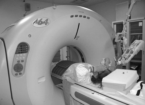
Multidetector computed tomography (MDCT) scans was performed to detect VTE.
Incidence of VTE after SHF was 18.1% and higher than the other operations. Advanced age, vessel damage by trauma, preoperative waiting time of bed rest may affect the high incidence rate. In this study, Chemoprophylaxis therapy was relatively effective in SHF (P=0.07). But anemia, hepatic dysfunction, increase of infection rate was reported after chemoprophylaxis therapy. Cautious use of chemoprophylaxis should be needed especially for an elder patient.
We examined 768 of 820 patients by MDCT. Routine screening with MDCT is controversial. MDCT is an invasive examination using contract media. It has been associated with risk of allergic reaction. However, the reliability of ultrasound in assessing VTE depends on the experience of the investigator, especially when assessing calf vein [14]. Uehara et al. reported that VTE was not revealed by ultrasound among the 2 of 6 subjects in whom VTE was detected in the lower extremities after TKA by MDCT [15]. They concluded that MDCT was the most sensitive modality. But the disadvantage of MDCT is that contrast media is needed as in venography in addition to radiation exposure. They note that this should be taken into account by physicians. We obtained informed consent from the all patients before MDCT was performed. On patient who had an allergy to the contrast media or refused to be examined by MDCT, Ultrasound was performed for detecting VTE by an experienced radiologist.
In conclusion, this study provides precise information by MDCT and Ultrasound on incidence for VTE in patients undergoing hip surgeries who were treated by almost same protocol at a single institution. The results of this data seem to indicate that the incidence of VTE after periacetabular osteotomy is significantly lower than surgery for hip fracture and relatively lower than THA and Revision THA. A younger age and non-invasion of the bone marrow of the femur may have affected the result. Chemoprophylaxis therapy was relatively effective especially on surgery for hip fracture.
CONFLICT OF INTEREST
The authors confirm that this article content has no conflict of interest.
ACKNOWLEDGEMENTS
The authors thank Hiromasa Tanino M.D., Tatsuya Sato M.D., Yasuhiro Nishida M.D. and Jane Weinhaus for their help.

