All published articles of this journal are available on ScienceDirect.

Traumatic Rupture of the Distal Triceps Tendon (A Series of 7 Cases)
Authors Info & Affiliations
Abstract
Even non-traumatic ruptures of the triceps tendon are rare, surgical therapy should be recommended in all cases, because of poor results after non-operative treatment. A golden standard for the surgical procedure is not established. A small series of traumatic distal tendon ruptures was treated surgical in our hospital and was followed up after 12 months concerning their function. Very good and good results could be found with a strong reintegration of the tendon by using transosseus sutures with non resorbable suture material. The refixation with suture anchors showed disappointing results with early pull-outs of the anchor. Revision with screw augmentation with a washer had to be performed. Concerning the biomechanical forces, which show up on the olecranon (up to 40 NM), the refixation of the triceps tendon has proved to be extremely resistant against pull out forces. The good results by using non absorbable transosseus sutures led to a standardized procedure in our trauma center, even the rupture is not traumatic.
INTRODUCTION
Ruptures of the distal triceps tendon (DTTR) are rare injuries [1]. DTTR are often found in combination with other injuries of the elbow or other joints [2, 3]. Recent studies on tendon avulsions reported of a frequency of 2% of DTTR [4]. However studies analyzing DTTR are rare [5]. DTTR have been reported to be associated with anabolic steroid use, weight lifting, and laceration. Other local and systemic risk factors include local steroid injection, olecranon bursitis, and hyperparathyroidism [6]. Also comorbidities such as renal failure, diabetes and rheumatoid arthritis can increase the risk for DTTR [7, 8]. In cases of secondary hyper- parathyroidism the osteoclastic activity is raised and often leads to dystrophic calcification weakening the tendon [9].
Often an indirect trauma by falling on the outstretched hand leads to distal triceps tendon injuries [10]. Thus an eccentrically overload of the contracted triceps muscle leads to the rupture [11, 12]. However, also direct force impact on the back on the elbow by falling on the flexed elbow has been reported in cases of triceps tendon ruptures too [12].
The typical injury of the triceps tendon is an avulsion from the osseous anchor of the tendon on the olecranon (Fig. 1). The diagnosis of distal triceps tendon ruptures is often difficult, so cases with misdiagnosed and untreated DTTR have been reported. In these cases a more complex surgical reconstruction has to be performed [13].
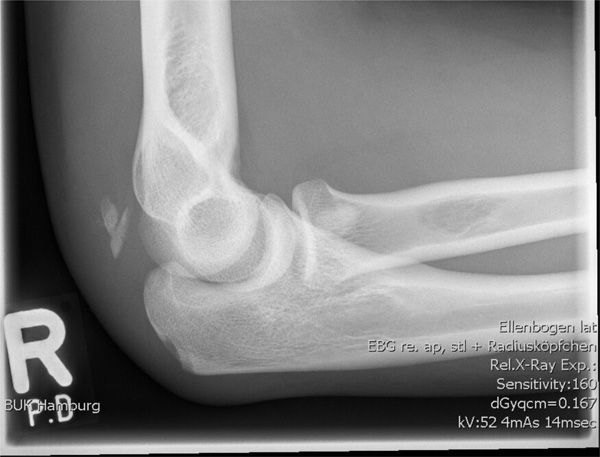
Typical Radograph of fresh distal triceps tendon rupture with dislocation of the osseus anchorage of the tendon.
Refixation of the triceps tendon rupture by transosseus sutures.

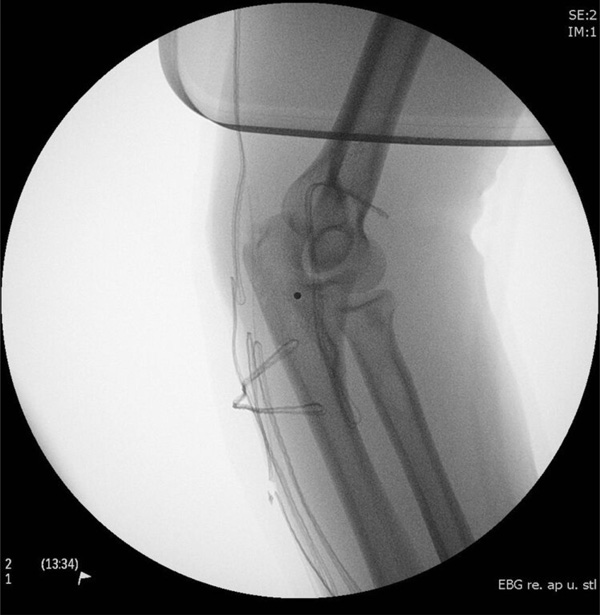
Dislocation of the suture anchor after refixation the triceps tendon rupture.
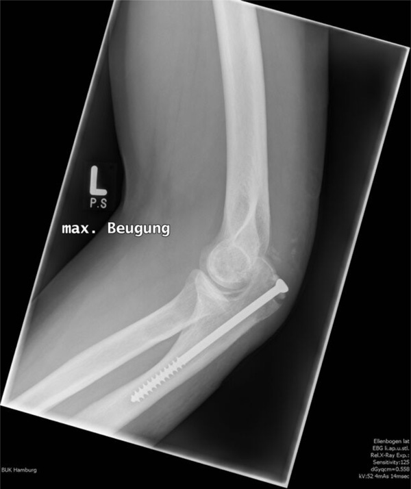
Radiograph after refixation the triceps tendon with transosseous suture and screw augmentation after suture anchor failure.
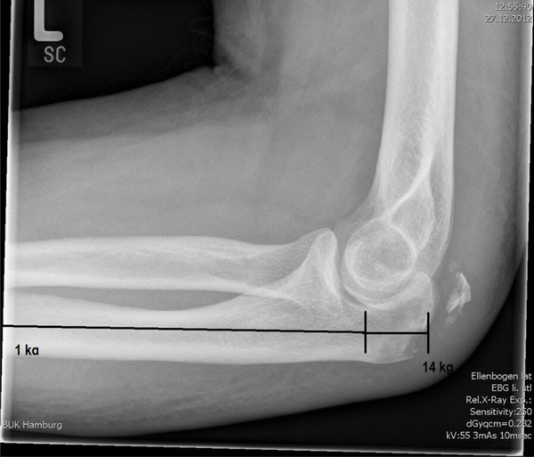
Approximated calculation of lever forces on the olecranon, when loading the distal ulna with 1kg of weight.
Number of cases according Morrey score.
| Morrey-Score | 100-95: very good | 95-85: good | 80-50: moderate | <50: bad |
|---|---|---|---|---|
| 6 | 1 | None | None |
Number of cases according Radin and Riseborough score.
| Radin and Riseborough Score | Good | Moderate | Bad |
|---|---|---|---|
| 6 | 1 | None |
Morrey score.
| Points | |
|---|---|
| Pain | |
| No pain | 30 |
| little pain, no pain killer | 25 |
| little pain, seldom pain killer | 15 |
| strong pain, frequent pain killer | 10 |
| strong and constant pain | 5 |
| unusable | 0 |
| Force 5: normal; 4: well; 3: moderate; 2: bad; 1: minimal; 0: paralytic | |
| Flexion | max. 5 |
| Extension | max. 4 |
| Pronation | max. 3 |
| Supination | max. 3 |
| Range of Motion | |
| Flexion | |
| 30° | 0 |
| 30° - 50° | 3 |
| 50° - 70° | 6 |
| 70° - 90° | 9 |
| 90° - 100° | 11 |
| 100° - 110° | 13 |
| 110° - 120° | 15 |
| > 120° | 17 |
| Extension | |
| 10° | 8 |
| 10° - 30° | 7 |
| 30° - 50° | 5 |
| 50° - 70° | 3 |
| 70° - 90° | 0 |
| Pronation / Supination | |
| 0.1 points per degree | max. 6 |
| Instability | |
| Anterior/posterior | |
| none | 3 |
| little < 5mm | 2 |
| moderate < 10mm | 1 |
| severe > 10mm | 0 |
| Medial/Lateral | |
| none | 3 |
| little < 5mm | 2 |
| moderate < 10mm | 1 |
| severe > 10mm | 0 |
| Function 1: normal; 0.75: moderate; 0.5: bad; 0.25: with help; 0: impossible | |
| Use back pocket | |
| Rise from chair | |
| Perineal care | |
| Wash opposite axilla | |
| Eat with utensils | |
| Comb hair | |
| Carrying 5-7kg | |
| Dress | |
| Pulling | |
| Throwing | |
| Do usual work | |
| Do usual sport sports | |
| Subjective assessment | |
| Very good | 3 |
| good | 2 |
| moderate | 1 |
| bad | 0 |
| Maximum possible points | 100 |
Radin and Riseborough score.
| Good | Loss of ROM < 10° in all direction, no pain |
| Moderate | Loss of ROM < 30° in all directions and/or little pain |
| Bad | Loss of ROM > 30° in all directions and/or constant pain |
Patients with DTTR often present with a swollen elbow due to hematoma and the disability to extend the flexed elbow against force. Fractures of the olecranon can be detected by conventional radiography. In addition, in clinically unclear cases, DTTR can be detected by MRI or sonography [14, 15].
Studies on cases of missed diagnosis or delayed therapy often report poor results with pain and loss of power of the extension of the elbow [16, 17].
The treatment of choice is surgical and often performed by using nonabsorbable sutures via drill holes in the olecranon for refixation (Fig. 2) [11]. Other studies also reported good results by using suture anchors. Arthroscopic treatment of DTTR has been reported in case reports or in treatment of partial avulsion of the triceps tendon as well [18, 19].
However a standard technique for the surgical approach of the rupture of the triceps tendon has yet to be established [20].
Thus, we analyzed our patients with traumatic triceps tendon ruptures in regards to their best operative treatment and clinical outcome.
MATERIAL AND METHODS
Seven patients (5 male, 2 female) with DTTR were treated surgically in the BG Trauma Center Hamburg between January 2011 and December 2012. All these patients were included, who showed up in the reported time period with a traumatic DTTR, reporting of posterior hit of their elbows. Exclusion criteria were non-traumatic DTTR and cases with concomitant elbow injuries. All patients included in this study provided written Informed Consent and met all inclusion criteria.
The mean age was 49 years ± 18 years. The mean time of follow up after surgery was 12 months ± 1.5 months.
All patients in this study had a direct contact trauma by falling on or hitting a fixed resistance with the posterior elbow. No comorbidities which could affect the integrity of the triceps tendon were detected. All patients were examined clinically and conventional radiographs were obtained.
The treatment was surgical in all cases. All operations were performed by two senior physicians. Refixation of the triceps tendon was performed by nonabsorbable sutures through bone tunnels in 6 cases. In one patient refixation of the tendon was accomplished by suture anchors (FASTak®,
Arthrex GmbH, Munich, Germany). The postoperative procedure included an immobilization by a cast in outstretched position. Passive flexion up to 30° of elbow flexion was allowed for the first two weeks and was increased every two weeks by 30° degrees. After 6 weeks postoperatively no further movement restrictions were applied and the cast was removed.
The clinical examination included evaluation of the Morrey Score (Table 3) and Radin and Riseborough score (Table 4) [21, 22]. The Morrey score was initially developed to access the function and pain level after injuries of the radial head and includes especially several functional tests to evaluate the range of motion of the elbow. The Radin and Riseborough score was developed for bony injuries of the elbow as well and represents a score, which includes pain und function and is easy in its application. The advantage of these both scores lies in their specification to access particular functional disorders and pain of the elbow. The subjective evaluation of the clinical outcome of the patients elbow function was assessed by the DASH score (disability of arm, shoulder and hand) [23]. The radiological follow-up was performed by a conventional radiograph of the elbow respecting the often visible osseous avulsion of the triceps tendon.
RESULTS
At follow-up of all cases were presented with a secure engraftment of the triceps tendon. Infections or soft tissue complications were not detected. In regards to the Morrey score 6 patients showed a very good result; one patient had a good result (Table 1). In regards to the Radin and Riseborough score, 6 patients demonstrated a good result, one patient showed only a moderate result (Table 2). The mean result in the subjective DASH score was 10.3. At follow-up radiological assessment showed no dislocation of the refixated avulsion of the triceps tendon or further calcifications. No patient had neurologic disorders on the treated extremity. All patients showed full extension of the elbow in the follow-up examinations. One patient had small loss of flexion (110°). All other showed good results concerning the flexion of the elbow (>120°). The rotation of the forearms of all patients was not restricted.
One patient treated with suture anchors needed an early revision of the suture because the suture anchor was pulled out in the postoperative radiograph (Fig. 3). This patient was then treated with conventional nonabsorbable sutures and screw augmentation (Fig. 4). Follow-up radiographs showed no dislocation of the originally dislocated osseous anchor of the triceps tendon. Besides no further complications have been observed.
DISCUSSION
Non-traumatic ruptures of the distal triceps tendon are an extremely rare injury (0,8% of 1014 tendon injuries) with no gold therapeutic standard [1]. We present our experiences in the treatment of isolated traumatic triceps tendon injuries, with a small group of 7 patients followed up. Statistical significant results cannot be expected due to the small number of patients and the rarity of this injury. But the bad results expectable after conservative treatment should at least be worth a try to evaluate the best therapeutic method for this injury.
All surgically treated tendon injuries were included into this assessment. In all cases presented, a solid refixation of the triceps tendon was achieved. The clinical results overall showed very good to good results with a high satisfactory rate among our patients. Concerning the pulling out of the suture anchor in one case, the subsequently performed operation by strong nonabsorbable sutures lead to a good result. Concerning the daily use of the elbow we achieved a good quality of live in our patients with the above documented results concerning flexion and extension of the elbow. Furthermore the results are convincing, if one considers the normal range of elbow motion in flexion and extension is approximately 0° to 140°, with a range of 30° to 130° required for most activities of daily living [24-26].
The comparison of our results with the literature is difficult, due to often published case reports, which miss usually a scoring examination. Heikenfeld et al. examined superficial, partial injuries of the triceps tendon and could show similar result according the DASH score (15.6) [19]. To our best knowledge the Morrey score und the Radin and Risbourogh score have not been applied to series of triceps injuries, but studies concerning evaluation of other elbow injuries (radial head, olecranon) use these scores regularly [27-29].
Due to the mandate of our trauma center to treat mainly work-related accidents, a number of patients who were initially treated in local centers could be examined afterwards in our clinic. Thus we were able to observe further patients with this specific injury, who were however not included into this study. By that we could compare different methods of refixation for the triceps tendon and saw cases with failure or secondary dislocation of the refixation, when suture anchors or small screw fixation was performed. Due to the complications in treatment of olecranon fractures or olecranon osteotomies the fixation of these fractures and their union can be complicated [30, 31]. The difficulty of refixation by suture anchors becomes clear if the direction of tensile forces on the anchor is considered. Only in 90° degree flexion of the elbow, the implanted suture anchors at the footprint of the triceps tendon can perform their maximum anchoring force. Clevenger et al. could demonstrate a significant lower anchoring force of suture anchors, if the tensile force to the anchor is below 90° degree (outstretched elbow) [32].
Transferring the lever principle to the forearm visualizes high forces taking effect on the footprint of the triceps tendon at the tip of the olecranon. Approximately 12-14 kg of weight take effect on the olecranon if you load the distal ulna with only 1 kg of weight (Fig. 5). According to these experiences, refixation of the triceps tendon needs to withstand high pull out forces and can be difficult. Nikolaidou et al. report in their case study extension forces up to 40Nm in bodybuilders [33].
In our opinion suture anchors are not strong enough to withstand these loads of tension in different bending degrees of the elbow, although some authors published promising results [20].
Some studies report the use of additional autograft in top- level athletes [34, 35], or even synthetic augmentation in cases of poor tendon qualities [6, 13]. To our knowledge no comparative studies using augmentation techniques with allografts versus autografts have been published, so the efficiency of the augmentation technique cannot be fully evaluated.
These experiences concerning the different refixation methods for distal triceps tendon ruptures led to standardized treatment of these injuries in our trauma center. All patients who show up the first time with a distal triceps tendon rupture are treated surgically by strong nonabsorbable transosseus sutures. In case of revision a further augmentation with screw and washer will be performed.
CONFLICT OF INTEREST
The authors confirm that this article content has no conflict of interest.
ACKNOWLEDGEMENTS
Declared none.

