All published articles of this journal are available on ScienceDirect.

Total Knee Arthroplasty for Rheumatoid Arthritis with mild extra-articular femoral anterocurvatum deformity: A Case Report and Literature Review
 Ryujiro Ohkado1
Michio Shinohara1
Ryutaro Kuriyama1
Tetsu Tsubone1
Koichiro Ihara1
Kazuhiro Yamazaki1
Ryujiro Ohkado1
Michio Shinohara1
Ryutaro Kuriyama1
Tetsu Tsubone1
Koichiro Ihara1
Kazuhiro Yamazaki1
Authors Info & Affiliations
Abstract
Background
Total knee arthroplasty (TKA) has achieved excellent outcomes through accurate bone resection and soft tissue balancing. TKA in patients with osteoarthritis (OA) or rheumatoid arthritis (RA) with extra-articular femoral deformity is technically difficult. Femoral deformity correction and TKA are often necessary in such cases.
Case Presentation
A 63-year-old woman had also been diagnosed with RA 18 years earlier. Her RA was relatively well controlled. The patient presented with severe right knee pain and gait disturbance. The patient had femoral exostosis and underwent operative therapy (resection) 47 years earlier. Radiographic evaluation revealed Larsen grade 4 arthritis of the knee joint and 8° femoral anterocurvatum deformity, measured relative to the anatomical axis of the femur. Standard TKA was performed with intra-articular correction. An anterior femoral notch (AFN) with 5.4mm was identified intraoperatively. 12 months postoperatively, the symptoms disappeared. The anterior femoral notch depth decreased to 2.8mm.
Conclusion
Standard TKA with intra-articular correction can provide satisfactory clinical short-term outcomes in patients with mild extra-articular femoral anterocurvatum deformity when appropriate preoperative planning and surgical technique are applied.
1. INTRODUCTION
Standard Total knee arthroplasty (TKA) is an excellent surgical procedure and has achieved good results. However, TKA for knee osteoarthritis (OA) or rheumatoid arthritis (RA) with extra-articular deformity is technically more challenging than standard TKA [1-5]. Previous studies have suggested that intra-articular resections combined with proper soft-tissue tensioning may be used to correct extra-articular angular deformities of up to 20° in the femur and 30° in the tibia [3, 6-9]. Nevertheless, the decision-making process becomes more challenging in patients with RA, in whom bone quality may be compromised due to systemic inflammation and long-term pharmacological treatment. In addition, a history of prior femoral surgery may further complicate anatomical alignment and influence surgical planning.
Most existing reports have focused on coronal-plane deformities, whereas relatively few studies have examined sagittal-plane deformities [10-12]. Moreover, postoperative changes in anterior femoral notch (AFN) morphology and their clinical implications remain insufficiently characterized [13, 14].
The present case is of particularly clinical interest because it illustrates surgical decision-making in an RA patient with a history of femoral surgery, demonstrates the feasibility of intra-articular correction for a mild sagittal femoral deformity, and provides insight into the postoperative evolution of an AFN.
We hypothesized that mild sagittal extra-articular femoral deformity in an RA patient could be adequately corrected by intra-articular resection during standard TKA without the need for additional femoral osteotomy, while maintaining satisfactory alignment and soft-tissue balance.
2. CASE REPORT
A 63-year-old female presented with severe right knee pain and gait disturbance. She had undergone surgical resection of a femoral exostosis 47 years earlier and had remained asymptomatic thereafter. She also worked as a caregiver. She had been diagnosed with RA 18 years prior and was being treated with methotrexate and adalimumab, with good disease control. However, she experienced faint pain in the left knee and had undergone left TKA eight years previously, with good post-operative outcome. However, right knee pain had been increasing over the past one year. Conservative treatment at a nearby hospital failed to provide relief, and she was referred to our department for further surgery.
Preoperative clinical assessment revealed a DAS28-CRP score of 2.43 indicating low RA disease activity, a C-reactive protein level of 0.05 mg/dL. The Hospital for Special Surgery (HSS) score was 52. The patient was 154cm tall and weighted 71kg, corresponding to a body mass index (BMI) of 31.6. Physical examination revealed swelling and tenderness in the right knee. The range of motion (ROM) was limited to 13 to 90°. The patellofemoral joint showed no abnormalities. Lower limb muscle weakness was not significant. No leg-length discrepancy was observed.
Radiographic evaluation demonstrated advanced degenerative changes consistent with Larsen grade 4 arthritis in the right knee joint (Fig. 1A). Lateral radiographs revealed an 8° femoral anterocurvatum deformity, measured relative to the anatomical axis of the femur (Fig. 1B). A weight-bearing anteroposterior radiograph revealed that the lateral distal femoral angle was 85°, and the anatomical femorotibial angle (FTA) was 183° (Fig. 2).
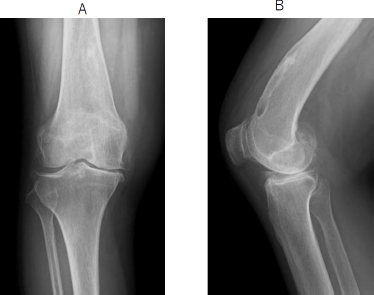
Preoperative radiograph of the right knee.
A Anterior-Posterior view.
B Lateral view demonstrating 8° sagittal femoral anterocurvatum deformity, measured relative to the femoral anatomical axis.
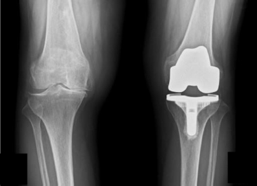
A weight-bearing anteroposterior radiograph demonstrating advanced rheumatoid arthritic changes (Larsen grade 4)
2.1. Surgical Procedure
TKA was performed under general anesthesia using a cemented posterior-stabilized prosthesis (Initia PS; Kyocera Co., Japan) based on a mechanical alignment strategy. The patella was replaced. Given the mild magnitude of the deformity and the absence of significant coronal malalignment, an intra-articular correction approach was selected, and no femoral osteotomy was planned. To address the sagittal deformity, the femoral component was placed in slight flexion (Fig. 3A). During surgery, the entry point for the intramedullary alignment rod was adjusted anteriorly to minimize the influence of the deformity. Distal femoral resection was performed at a depth of 8.5mm from the most distal condyle. The proximal tibial resection depth was 9mm from the lateral tibial plateau. Detailed intraoperative records regarding the exact degree of flexion used for femoral component placement were unavailable because the procedure was performed before initiation of this study. Gap-balancing technique was used to achieve appropriate ligament tension. No additional soft-tissue release was required, and balanced flexion and extension gaps were obtained. Patellar resurfacing was performed because patients with RA are reported to have a higher prevalence of patellofemoral joint involvement and anterior knee pain. A size 7 femoral component and size t tibial component were implanted. The total operative time was 114 minutes.
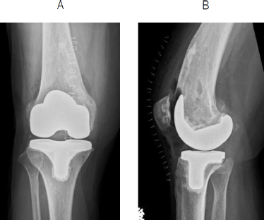
Postoperative radiograph.
A Anterior-posterior view.
B Lateral view demonstrating an anterior femoral notch with a depth of 5.4mm.
An anterior femoral notch was identified intraoperatively, measuring 5.4 mm in depth and 9 mm in length (Fig. 3B).
Postoperatively, ROM exercise and partial weight bearing was initiated, following by progression to full weight-bearing by six weeks. The postoperative course was uneventful. At the 12-month follow-up, the patient reported complete resolution of pain. The ROM improved to 0–120°, and the HSS score increased to 92, exceeding the minimal clinically important difference. No anterior knee pain was reported.
Radiographic imaging showed that the depth of the AFN decreased from 5.4mm immediately postoperatively to 2.8mm at follow-up (Fig. 4).
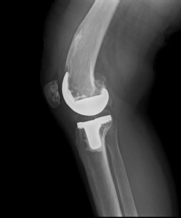
Postoperative radiograph at 12 months postoperatively.
Lateral view.
The anterior femoral notch depth decreased to 2.8mm.
3. DISCUSSION
Extra-articular knee deformities are commonly caused by a previous traumatic event, metabolic bone disease, stress fracture, or previous surgical intervention [8, 13]. In the present case, the deformity resulted from a previous surgical intervention for femoral exostosis. TKA for severe osteoarthritis associated with severe extra-articular deformity has been reported to be technically challenging. Correction of the alignment of the lower extremity can be accomplished by osteotomy of either the distal part of the femur or the proximal tibia [1-5]. When performing TKA, extra-articular deformities have to be considered to achieve satisfactory ligament balance, acceptable component alignment, and better restoration of the mechanical axis in both the coronal and sagittal planes [5]. For severe deformities, corrective osteotomy combined with TKA may be necessary; however, this approach is technically demanding and has been associated with complications such as delayed union or nonunion [8, 15].
In contrast, intra-articular correction may be appropriate for mild deformities when adequate ligament balance can be maintained [6, 7]. Wallace et al. emphasized that excessive asymmetric bone resection may compromise stability, whereas Sculco et al. warned that overcorrection can lead to flexion-extension gap imbalance [16, 17].
While previous studies have proposed criteria for selecting intra-articular correction or corrective osteotomy, there remains no consensus regarding the optimal management of extra-articular deformities around the knee [7, 18].
While intra-articular correction is well established for coronal deformities, there is relatively limited literature addressing sagittal deformities [10-12]. In the present case, the deformity was limited to 8° in the sagittal plane, with no substantial coronal malalignment. Therefore, intra-articular correction was considered sufficient and allowed satisfactory alignment and soft-tissue balance without the need for additional femoral osteotomy.
The AFN is a well-known complication of TKA. Concerns exist regarding the impact of the anterior femoral notch on knee ROM and anterior knee pain, and the potential risk of postoperative periprosthetic fractures. Benkovich et al. reported anterior femoral notching compromises femoral integrity, with defects>2.5mm significantly increasing fracture risk [14]. Puranik et al. reported that there is no correlation between notching and supracondylar fracture of the femur following TKA. It does not appear to have any bearing on the range of movement or outcome scores [19]. In the present case, no fracture or anterior knee pain was observed. Interestingly, AFN depth decreased from 5.4 mm to 2.8 mm during follow-up. The observed decrease in AFN depth should be interpreted cautiously because measurements obtained from two-dimensional lateral radiographs are susceptible to projectional variation. Three-dimensional CT assessment would provide more accurate quantification of notch morphology and should be considered in future studies. Furthermore, bone metabolism in RA patients may differ from that in patients with osteoarthritis, potentially influencing the remodeling process.
Recent advances in robotic-assisted TKA have improved the accuracy of bone resection and implant positioning, particularly in complex deformities [20, 21]. Robotic systems may allow more individualized adjustment of sagittal femoral resection angles in patients with femoral bowing deformities. Nevertheless, the present case demonstrates that satisfactory clinical and radiographic outcomes can also be achieved using conventional instrumentation when careful preoperative planning and meticulous surgical technique are employed.
4. STUDY LIMITATIONS
This report describes a single case and therefore has limited generalizability. The follow-up period was relatively short. AFN measurements were performed using two-dimensional radiographs and may have been affected by projectional variability and measurement error. Three-dimensional imaging modalities such as CT may provide more accurate assessment of AFN morphology. Furthermore, no biochemical markers of bone turnover were evaluated, preventing objective assessment of postoperative bone remodeling.
CONCLUSION
Standard TKA with intra-articular correction achieved satisfactory clinical and radiographic outcome in this patient with a mild extra-articular deformity. Careful preoperative planning and precise component positioning are essential to maintain proper alignment and soft-tissue balance. In selected cases, intra-articular correction may be a viable option for minor deformities, potentially avoiding the need for additional osteotomy. Intraoperative mild anterior femoral notch defects may undergo postoperative bone remodeling, although the clinical significance of this finding remains unclear.
AUTHORS CONTRIBUTION
The authors confirm their contribution to the paper as follows: T.M.: Designed the study, analyzed the data, and wrote the manuscript; M.S., R.K., T.T., K.I. and K.Y.: Collected the data and participated in the design of the study; R.O.: Analyzed the data, and helped write. All authors reviewed the results and approved the final version of the manuscript.
LIST OF ABBREVIATIONS
| TKA | = total knee arthroplasty |
| OA | = osteoarthritis |
| RA | = rheumatoid arthritis |
| AFN | = anterior femoral notch |
| HSS | = Hospital for Special Surgery |
| ROM | = range of motion |
ETHIC APPROVAL AND CONSENT TO PARTICIPATE
Institutional ethics committee approval was not required for this retrospective single-patient case report.
HUMAN AND ANIMAL RIGHTS
All procedures performed in studies involving human participants were in accordance with the ethical standards of institutional and/or research committee and with the 1975 Declaration of Helsinki, as revised in 2013.
AVAILABILITY OF DATA AND MATERIAL
Data are available from the corresponding author upon reasonable request.
ACKNOWLEDGEMENTS
Declared none.