All published articles of this journal are available on ScienceDirect.

Kinematics and Electromyographic Analysis of Three Types of Internal Rotation in the Shoulder
 iD
Hiroshi Karasuno3
iD
Kunio Yoshizaki4
iD
Kazuhiro Endo1
iD
Ryo Sahara1
iD
iD
Hiroshi Karasuno3
iD
Kunio Yoshizaki4
iD
Kazuhiro Endo1
iD
Ryo Sahara1
iD
Authors Info & Affiliations
Abstract
Introduction
Kinematic and electromyography (EMG) studies concerning internal rotation (IR) of the shoulder are limited. This study investigated the kinematic components and EMG activity of the rotator cuff muscles during IR at the side (IRs), IR at 90 ° of abduction (IRa), and IR to the posterior (IRp).
Methods
Ten healthy young men attended this study. The angles of IR, extension, and abduction of the humerothoracic joint, and angles of scapular IR, anterior tilt, and upward rotation were measured using a three-dimensional (3-D) motion analyzer. EMG activities in the 4 rotator cuff muscles were simultaneously recorded using synchronized 3-D motion analysis.
Results
The maximum IR angles were 56.6°, 72.4°, and 31.6° in IRs, IRa, and IRp, respectively. The maximum extension angle was 34.1° in IRp, and the maximum abduction angles were 1.4°, 87.4°, and 24.1° in IRs, IRa, and IRp, respectively. The EMG activity patterns of the rotator cuff muscles differed across the 3 IR types. %IEMGs of the subscapularis in IRs were approximately 20%, gradually increased up to 18.2% in IRa, and greatly increased to 29.8% at maximum IRp. %IEMGs of the supraspinatus were constant during the 3 movements, and EMG activities of the infraspinatus and teres minor decreased preceding IRa, whereas those increased up to maximum IRp.
Discussion
The rotation axes of IRs and IRa are constant, but that of IRp always changes. Our results show that the internal rotation angles for IRs and IRa are nearly identical to those reported previously. In contrast, the rotation angle for IRp differs from those in the current reports. EMG analysis of the 3 types of IR movements reveals different muscle activity patterns.
Conclusion
Kinematic and EMG analyses revealed that the three types of IR completely differ. The total IR angles in the humerothoracic joint increased in the order IRa > IRs > IRp. The highest activity of the subscapularis was observed in IRp. The internal rotation angle is the smallest, and the internal rotation movement of the IRp is completed in the early phase of the movement. The joint structure itself is presumed to limit the internal rotation movement of the IRp.
1. INTRODUCTION
The range of motion (ROM) of the shoulder and elbow joints is crucial for upper limb function and is related to the ability to perform activities of daily living (ADLs). Two ADLs, including combing hair and washing the back, correlate significantly with shoulder external and internal rotation [1]. Studies on the relationship between shoulder ROM and ADLs have shown a low correlation between external shoulder rotation and ADLs, whereas internal shoulder rotation (IR) is highly correlated with ADLs [1, 2]. Shoulder IR is an essential motion for several ADLs: IR to the posterior (IRp) needs to reach the seventh thoracic vertebra to wipe the bottom on a toilet, put on a bra, fasten bands into pants, or take a wallet from a back pocket. Active IR loss is commonly observed in patients with rotator cuff tears, frozen shoulders, and those who undergo reverse shoulder arthroplasty [2-5]. The inability to perform IRp severely limits shoulder function, and patients experience a greater negative impact than expected when considering IRp under the first lumbar spine [2, 6].
Physicians commonly measure three types of shoulder IR in patients with IR deficits: IR at the side (IRs), IR at 90° of abduction (IRa), and IRp. IRp and IRa were moderately limited in patients with rotator cuff tears; however, IRs were not limited, and the angles of IRs before and after treatment were not significantly different [7]. Although mild IRs and severe IRp loss have been observed in patients with refractory frozen shoulder [8, 9], the discrepancy between IRs and IRp deficits has been difficult to explain. Moreover, the reason why the IRp deficit is refractory to improvement, unlike shoulder elevation and external rotation, has not been clarified [10]. The etiology of IRp deficit has been explained by the posteroinferior capsular thickening and tightness of the supraspinatus, infraspinatus, and teres minor [11, 12]. Stretching and arthroscopic capsular release have been proposed as effective approaches for IRp deficit [13-15]. Understanding the pathophysiology and providing appropriate treatment options for IRp deficit remains challenging.
Many kinematic and electromyographic studies have been conducted on shoulder elevation, whereas few kinematic papers on shoulder IR movements have been published. Mallon et al. [16] indicated that the IR angle of the glenohumeral joint was 20° in maximum IRs and 60° in IRp at the highest vertebral level and that the maximum IRp occurred in a 2:1 ratio between the glenohumeral and scapulothoracic joints. The IR angle of the glenohumeral joint was approximately 70°, and the angle of elbow flexion was 100° in the IRp up to the sixth thoracic vertebra level [17]. To date, only two studies on IRa movement of the shoulder joint have been found [18, 19]; however, comparative studies of the three types of shoulder IR are lacking. This study aimed to measure kinematic components of the humerothoracic and scapulothoracic joints and to analyze electromyographic activity of the rotator cuff muscles during IRs, IRa, and IRp. We hypothesized that the kinematic components and activity patterns of the rotator cuff muscles would be different among the IRs, IRa, and IRp.
2. METHODS
2.1. Participants
This study included 13 male university students with no history of shoulder pain or injury who had not participated in throwing sports. Examinations of muscle atrophy, scoliosis, scapular dyskinesia, general joint laxity, instability, shoulder ROM, impingement tests, and plain X-ray of their shoulder joints were performed. Three of them, observing shoulder abduction restriction, scapular dyskinesia during shoulder elevation, or positive instability tests, were excluded. Finally, the remaining 10 individuals, who demonstrated no abnormalities on examination and had normal radiographs, were included in this study (mean age: 21.5 ± 3.4 years, mean height: 1.72 ± 0.12 m, mean body weight: 65.8 ± 7.1 kg, and mean body mass index: 21.6 ± 3.3). Calculations using G*Power 3.1 with an effect size of 0.8, α = 0.05, and power = 0.95 resulted in a total sample size of 10. As it was evident that the difference in mean values was substantial, a sample size of 10 was used. The right arm was tested as the dominant arm in all participants. Kinematics and electromyography during IRs, IRa, and IRp of the right shoulders were recorded. The Institutional Review Board of our hospital approved this study. All participants were informed of the nature and details of the study and provided written and verbal consent before their participation. The experiment ran from April to September 2021, and data analysis and interpretation were performed in 2022.
2.2. Shoulder Kinematic Measurements
Three-dimensional (3-D) biomechanical data of the shoulder joint were collected using a computerized motion analysis system (MAC 3D System, Motion Analysis Corp., Santa Rosa, CA, USA). This system enabled 50 Hz data acquisition from 10 synchronized infrared cameras positioned around the participants. Motion data were analyzed with the KineAnalyzer system software (Kissei Comtec Co., Ltd., Nagano, Japan). The skin displacement distance between each marker of the acromion angle, the scapular spine, anterior and posterior acromion angles, lateral and medial epicondyles, thoracic vertebrae, and other anatomical landmarks was measured by X-ray films and palpation to find the appropriate marker position that minimized skin displacement between the starting positions and maximum IRs.
Methods in this study were the same as those in our previous studies [20, 21]. The participants stood with their thoracic spines, arms, pelvis, and knees exposed. Reflective markers were attached to the anterior and posterior sides of the humeral head, the midpoint of the scapular spine, the lateral and medial epicondyles of the humerus, the styloid process of the radius, distal ulna, spinous processes of the 7th cervical spine, 7th thoracic spine, and 5th lumbar spine, the top and xiphoid process of the sternum, and bilateral anterosuperior spines of the pelvis. The line connecting the midpoint of the anterior and posterior markers of the humeral head to the midpoint of the medial and lateral epicondyles was considered the humeral axis. The scapular spine line was recognized as the line that delineated the midpoint of the scapular spine and the posterior acromion. The IR angle of the humerus was computed from the rotation angle of the line connecting the lateral and medial epicondyles. Every IR motion began from each initial position, and the starting position angles of the humerus, scapula, and thoracic spine were set at 0° [22]. The IR, abduction, and extension angles of humerothoracic movement and scapulothoracic rotation were simultaneously measured. The maximum value of each IR movement was set to 100%, and the mean IR angles were displayed in 20% increments. Every angle was autocorrected using the numeric value of the thoracic spine tilt angle.
2.3. Testing Protocol
The starting position of IRs was the arm at the side with the elbow flexed to 90 °, and participants internally rotated the arm until the forearm contacted the abdomen. IRa started at 90° of shoulder abduction and the neutral position of external and internal rotation with 90° of elbow flexion, and the arm reached maximum IRa. The IRp was initiated from the drooping position of the upper limb, the palm faced backwards, and the thumb directly reached the seventh thoracic vertebra. Participants practiced several times to complete three IR movements within 5 seconds before the measurements. The investigators were responsible for ensuring that the participants maintained proper arm position and motivation during each trial. After confirming that three IR movements were proper, they performed the IRs, IRa, and IRp movements three times in this order to collect data. A 3-minute rest was taken between the three types of internal rotation movements, and a 30-second rest was taken between repetitions of the same internal rotation movement.
2.4. Electromyography of Rotator Cuff Muscles
Simultaneous electromyographic (EMG) data were acquired from the supraspinatus, infraspinatus, subscapularis, and teres minor muscles, synchronized with 3-D motion analysis during three distinct IR movement patterns. We prepared fine wires for intramuscular use following previous reports [21, 23]. Two sterile 50-µm-diameter wires coated with Teflon (Dupont, Wilmington, DE, USA) were inserted into the belly of rotator cuff muscles using a 25-gauge hypodermic needle. First, we inserted a needle electrode into the supraspinatus, in the area bordered by the scapular spine, acromion, and clavicle; the needle tip was positioned 4 mm above the supraspinatus fossa. Next, a needle electrode was placed in the mid-portion of the infraspinatus muscle, using the same methodology as for the supraspinatus. A needle electrode was inserted into the inferior part of the subscapularis, which was identified as the primary internal rotator during IRs and IRa [24]. We inserted a needle electrode in the teres minor under ultrasound guidance. Participants performed shoulder joint movements to verify accurate electrode placement for each rotator cuff muscle.
EMG data were acquired using the EMG-100C system (BIOPAC Systems Inc., CA, USA), synchronized with a 3-D motion analysis system. Raw EMG signals were bandpass-filtered (10-1500 Hz) and root-mean-square smoothed (100 ms window). Subsequent data analysis was conducted using Bimutus 2 software (Kissei Comtec Co., Nagano, Japan) [21,25]. Maximum voluntary isometric contractions (MVIC) were performed for 5 seconds in standardized initial positions for IRs, IRa, and IRp (at the buttock level). EMGmax, defined as the average of three trials per second, served as the reference value for amplitude-normalized EMG. The integrated EMG (IEMG) of the rotator cuff muscles was calculated for each 15° increment. The percentage of IEMG (%IEMG) for each 15° increment per second was then determined from the average per-second values for each 100% MVIC across all participants [21].
2.5. Statistical Analysis
Statistical analyses were performed using SPSS Statistics 30.0 software (IBM Japan, Tokyo, Japan) with a significance threshold P-value < 0.05. All the data are shown as the mean ± standard deviation. The reliability of internal rotation, abduction, and extension of the humerothoracic joint, and internal rotation, upward rotation, and anterior tilt of the scapulothoracic joint during the three types of IR movements were evaluated with intraclass correlation coefficients (ICC [1,1] and ICC [1,3]). Data normality was assessed using the Shapiro-Wilk test. Tukey’s range test was adjusted for multiplicity using a simulation-based method, and adjusted P-values and 95% confidence intervals (95% CIs) were calculated. Tukey’s honestly significant difference test or Steel-Dwass test were performed to compare the angles of humerothoracic and scapulothoracic movements and the %IEMG of each rotator cuff muscle in 15° increments.
3. RESULTS
3.1. Repeatability of Humerothoracic and Scapulothoracic Joints
System measurement error was observed to be less than 0.05 mm for static length measurements, less than 0.28 mm for dynamic length measurements, less than 0.09° for static angular measurements, and less than 0.47° for dynamic angular measurements [21]. The reliability of the humerothoracic and scapulothoracic movements for the three IR movements is presented in Table 1. The ICC (1,1) of IR of IRp in the humerothoracic joint, IR of IRs, and upward rotation in the scapulothoracic joint of IRa showed 0.222, 0.279, and 0.456, respectively. . ICC (1,3) of IR of IRp in the humerothoracic joint demonstrated 0.461. The EMG reliability of rotator cuff muscles demonstrated no significant differences (P = 0.19 ‒ 0.86) [21].
| - | IRs | IRa | IRp | |||
|---|---|---|---|---|---|---|
| Humerothoracic Joint | ICC (1,1) | ICC (1,3) | ICC (1,1) | ICC (1,3) | ICC (1,1) | ICC (1,3) |
| Abduction | 0.858 (p < 0.001) | 0.948 (p < 0.001) | 0.572 (p < 0.005) | 0.801 (p < 0.005) | 0.891 (p < 0.001) | 0.961 (p < 0.001) |
| Extension | 0.630 (p < 0.001) | 0.836 (p < 0.001) | NA | NA | 0.849 (p < 0.001) | 0.961 (p < 0.001) |
| Internal rotation | 0.577 (p < 0.001) | 0.804 (p < 0.001) | 0.802 (p < 0.001) | 0.924 (p < 0.001) | 0.222 (p < 0.05) | 0.461 (p < 0.005) |
| Scapulothoracic joint | ICC (1,1) | ICC (1,3) | ICC (1,1) | ICC (1,3) | ICC (1,1) | ICC (1,3) |
| Internal rotation | 0.279 (p < 0.091) | 0.537 (p < 0.091) | 0.628 (p < 0.001) | 0.875 (p < 0.001) | 0.684 (p < 0.001) | 0.866 (p < 0.001) |
| Upward rotation | 0.675 (p < 0.001) | 0.537 (p < 0.001) | 0.456 (p < 0.05) | 0.716 (p < 0.05) | 0.853 (p < 0.001) | 0.946 (p < 0.001) |
| Anterior tilt | 0.700 (p < 0.001) | 0.875 (p < 0.001) | 0.949 (p < 0.001) | 0.982 (p < 0.001) | 0.766 (p < 0.001) | 0.908 (p < 0.001) |
Abbreviations: IRs: internal rotation at the side; IRa: internal rotation at 90º of abduction; IRp: internal rotation to the posterior; ICC: intraclass correlation coefficients; NA: not available.
3.2. Kinematic Elements of Three Types of Internal Rotation
The maximum movements of the humerothoracic joint, scapulothoracic joint, and trunk during three IR movements are shown in Table 2. The mean maximum angles of the IRs, IRa, and IRp in the humerothoracic joint were 56.6°, 72.4°, and 31.6°, respectively (Fig. 1). The mean maximum abduction angles of the IRs, IRa, and IRp were 1.4°, 87.4°, and 24.1°, respectively. The mean maximum extension angles were -8.1° in IRs and 34.1° in IRp. The scapular movements affecting the three types of IR were 6.4° of IR during IRs, 3.5° of upward rotation and 14° of anterior tilt during IRa, and 9.6° of anterior tilt during IRp. The trunk motions during the three IR movements showed a small number of angles.
| - | IRs (Degree) | IRa (Degree) | IRp (Degree) |
|---|---|---|---|
| Humerothoracic Joint | - | - | - |
| Internal rotation | 56.6 ± 8.7 | 72.4 ± 6.0 | 31.6 ± 6.8 |
| Abduction | 1.4 ± 3.6 | 87.4 ± 5.7 | 24.1 ± 8.0 |
| Extension | ‐8.1 ± 2.7 | NA | 34.1 ± 7.3 |
| Scapulothoracic joint | - | - | - |
| Internal rotation | 6.4 ± 2.8 | 3.5 ± 10.1 | 3.2 ± 2.9 |
| Anterior tilt | 2.2 ± 2.6 | 14.0 ± 9.9 | 9.6 ± 3.7 |
| Upward rotation | 1.8 ± 1.5 | 3.5 ± 3.6 | 4.1 ± 6.1 |
| Trunk | - | - | - |
| Flexion | 0.5 ± 0.4 | 0.7 ± 2.0 | 1.3 ± 0.9 |
| Lateral tilt | 0.1 ± 0.3 | 0.1 ± 0.6 | 0.1 ± 0.5 |
| Rotation | 0.5 ± 0.4 | 0.8 ± 1.2 | 0.3 ± 1.3 |
Abbreviations: IRs: internal rotation at the side; IRa: internal rotation at 90º of abduction; IRp: internal rotation to the posterior.
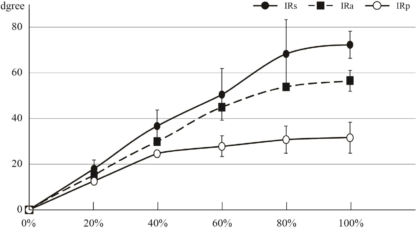
Internal rotation angles in three types of internal rotation of the shoulder.
IRs: internal rotation at the side; IRa: internal rotation at 90º of abduction; IRp: internal rotation to the posterior
3.3. Electromyographs of Rotator Cuff Muscles
Figure 2 and Table 3a, b, c illustrate the %IEMG activity of the rotator cuff muscles during the three types of IR. Distinct rotator cuff muscle activity patterns were observed across the three IR movements, and the %IEMG in every muscle was lower than 30%. The %IEMGs of the subscapularis in IRs were consistently 20%, gradually increased up to 18.2% in IRa, and greatly increased up to 29.8% at maximum IRp. The activity of the subscapularis in the IRp showed a statistically significant difference compared to that in the IRs (P < 0.01) and IRa (P < 0.01). The %IEMG of the infraspinatus gradually decreased, and that of the subscapularis was almost constant during IRs (Table 3a). The %IEMG of the infraspinatus gradually reduced, and that of the subscapularis increased in the progression of IRa (Table 3b). Interestingly, the %IEMG of all rotator cuff muscles increased as the hand position rose during IRp (Table 3c).
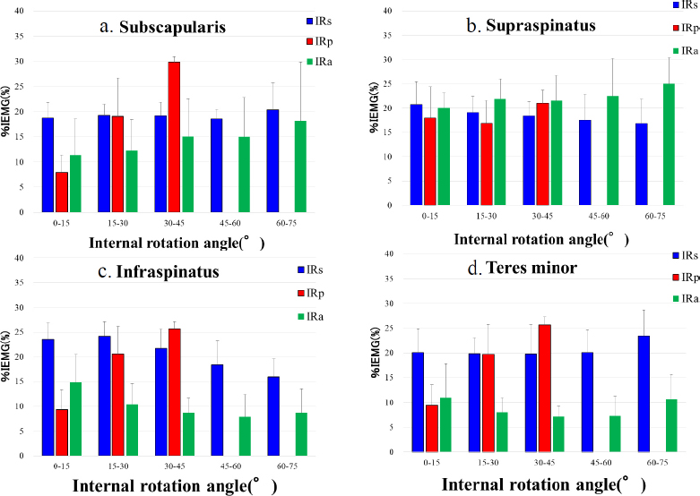
%IEMG of rotator cuff muscles during three internal rotations.
%IEMG: percent integrated electromyography
| Degree | 0-15 | 15-30 | - | 30-45 | 45-60 | 60-75 |
|---|---|---|---|---|---|---|
| Numbers | 10 | 10 | - | 10 | 10 | 10 |
| Supraspinatus (%) | 20.7 ± 4.7 | 19.1 ± 3.3 | ‡‡ | 18.4 ± 2.9 | 17.6 ± 5.2 | 16.9 ± 5.0 |
| Infraspinatus (%) | 23.6 ± 3.3 | 24.2 ± 2.9 | - | 21.7 ± 3.9 | 18.4 ± 4.8 | 15.9 ± 3.7 |
| Teres minor (%) | 20.1 ± 4.8 | 19.8 ± 3.2 | ‡ | 19.8 ± 5.9 | 20.1 ± 4.6 | 23.5 ± 5.2 |
| Subscapularis (%) | 18.8 ± 3.0 | 19.2 ± 2.2 | ‡ | 19.2 ± 2.7 | 18.6 ± 1.8 | 20.3 ± 5.4 |
Note: ‡p < 0.05, ‡‡p < 0.01 vs. infraspinatus
| Degree | 0-15 | - | 15-30 | - | 30-45 | - | 45-60 | - | 60-75 |
|---|---|---|---|---|---|---|---|---|---|
| Numbers | 10 | - | 10 | - | 10 | - | 10 | - | 20 |
| Supraspinatus (%) | 20.1 ± 3.1 | - | 22.0 ± 4.0 | - | 21.6 ± 5.2 | - | 22.5 ± 7.7 | - | 25.0 ± 5.4 |
| Infraspinatus (%) | 14.8 ± 5.6 | - | 10.4 ± 4.2 | ** | 8.7 ± 3.0 | **† | 7.9 ± 4.5 | ** | 8.7 ± 4.9 |
| Teres minor (%) | 11.0 ± 6.8 | * | 8.0 ± 3.0 | ** | 7.1 ± 2.1 | **† | 7.3 ± 3.9 | ** | 10.7 ± 4.8 |
| Subscapularis (%) | 11.3 ± 7.3 | * | 12.3 ± 6.1 | ** | 15.1 ± 7.5 | * | 15.0 ± 7.8 | - | 18.2 ± 11.7 |
Note: *p < 0.05, **p < 0.01 vs. supraspinatus; †p < 0.05 vs. subscapularis
| Degree | 0-15 | - | 15-30 | 30-45 | - |
|---|---|---|---|---|---|
| Numbers | 10 | - | 10 | 9 | - |
| Supraspinatus (%) | 18.0 ± 6.4 | - | 16.9 ± 4.7 | 21.0 ± 2.7 | †† |
| Infraspinatus (%) | 9.3 ± 4.1 | ** | 20.6 ± 5.6 | 25.7 ± 1.5 | †† |
| Teres minor (%) | 9.4 ± 4.2 | ** | 19.7 ± 6.0 | 25.7 ± 1.6 | †† |
| Subscapularis (%) | 8.0 ± 3.4 | ** | 19.0 ± 7.6 | 29.8 ± 1.1 | - |
Note: *p < 0.05, **p < 0.01 vs. supraspinatus; †p < 0.05, ††p < 0.01 vs. subscapularis.
4. DISCUSSION
The kinematic elements in this study indicated that the three types of shoulder IR were completely different movements. The total IR angles in the humerothoracic joint increased in the order IRa > IRs > IRp. Every scapular movement during the three types of IR ranged from less than 14°, which was much smaller than the 40° of scapular upward rotation during arm elevation [20, 21]. Therefore, scapular movements have a small influence on three IR movements compared with those during shoulder elevation. Since IRs is an internal rotation around the vertical axis of the humerus, and the axis of IRa is set at 90° of shoulder abduction, both IR movements are rotations around definite axes. In contrast, the rotational axis of the IRp varies with an increase in the abduction and extension angles of the humerothoracic joint. No EMG studies of IRp have been available, and this study is the first to compare the EMGs of the three types of IR movements. The activities of all rotator cuff muscles during IRs were almost constant, implying that IRs are an invariable movement of the humerothoracic joint. From the EMG data of the IRp, the muscle activity in the subscapularis, infraspinatus, and teres minor muscles continuously increased up to the maximum IRp, suggesting that IRp movement may be a complex motion.
The low intraclass correlation coefficient (ICC) was 0.222 for ICC (1, 1) and 0.461 for ICC (1, 3) of humerothoracic internal rotation of IRp. While the average internal rotation angle for IRp is 31.6 ± 6.8 degrees, it is about half that of IRs (56.6 ± 8.7) and IRa (72.4 ± 6.0), but the standard deviation values are almost the same. The decrease in ICC (1, 1) and ICC (1, 3) is considered to be due to the large variability in the average internal rotation angle of IRp. This indicates that internal rotation of the humerus finishes in the initial stages of IRp movement, and that IRp is then continued with movements such as extension of the humerus and anterior tilting of the scapula. The low ICC value of IRp movement indicates a significant individual difference. ICC (1,1) of scapulothoracic internal rotation for IRs was 0.279. Since the IRs indicate that the movement finishes when the forearm touches the abdomen, it is presumed that the humerus is not fully internally rotated. Internal rotation of the scapula is not required; therefore, the ICC (1, 1) becomes low. ICC (1,1) of scapulothoracic upward rotation for IRa was 0.456. The humerus abducts 90 degrees, and the scapula rotates upward in IRa. The humerus is maintained at 90 degrees of abduction, and the upward rotation of the scapula is also fixed. Because the internal rotation movement in IRa depends on the glenohumeral joint, this is presumed to be a factor in the low ICC (1, 1) value.
Several kinematic studies on the IR movements of the shoulder joint have been published: one study concerned IRs, two reported IRa, and two observed IRp. Mallon et al. [16] reported that the IR angle in the glenohumeral joint was 20° and that in the scapulothoracic joint was 2° at the maximum IRs, which were significantly smaller than our results of 56.6° and 6.4°. With the arm hanging down, retroversion of the humeral head ranges from 30° to 45° in the coronal plane [26]. As the retroversion and attachment of the subscapularis tendon to the lesser tuberosity affect the range of IR in the glenohumeral joint, the potential IR angle may range from approximately 45° to 60° in maximum IRs. The average IR angles of IRs pre- and post-intervention in patients with rotator cuff tears were described as 56.3° and 58.3°, respectively [7]; therefore, our results are considered reliable. In a preliminary study, we used computed tomography (CT) to measure the IRs angle in the supine position, finding that an increase in extension was accompanied by a decrease in the IR angle of the glenohumeral joint (data not shown). In Mallon’s study, the elbow appeared to touch the CT table in the supine position, and an increase in the extension angle of the glenohumeral joint may have decreased the IR angle of the joint.
Two studies showed kinematic data of the IRa: one study showed an IRa angle of 73° in the humerothoracic joint [18], and another study showed an IRa angle of 81° in the humerothoracic joint and an anterior tilt angle in the scapula of 16.5° [19]. Both IR and scapular anterior tilt angles in the studies coincided with our results of 72.4° and 14°, respectively; therefore, the IRa movement revealed high reproducibility. In contrast, the IRp is difficult to measure kinematically. The IR angles with the thumb at the highest vertebral level were described as 60° in the glenohumeral joint and 32° in the scapulothoracic joint during IRp using CT in the prone position [16]. Our study showed that the IR angles of the humerothoracic joint and scapulothoracic joint were 31.6° and 3.2°, respectively, at maximum IRp in the standing position, which were smaller than those in Mallon’s study. The IR angles of the glenohumeral and scapulothoracic joints in the prone position on the CT table were likely to increase. This position leads to IR of the scapula, bringing the elbow close to the CT table and further rotating the humerus internally. Therefore, the prone position provides larger IR angles in the glenohumeral and scapulothoracic joints compared to our results of IRp. Another study using an electromagnetic tracking device indicated that the IR angle of the shoulder joint was 73° from the hanging-arm position to the thumb at the 6th vertebra level [17]. The IR angle of the glenohumeral joint was more than twice that in our results. Our study differed from that study in the following three aspects: measurement devices, starting positions, and measurement angles. The starting position of the study was with the arm hanging down, 90° flexion of the elbow, and the thumb reaching the buttock, sacrum, and every vertebral level under static positions. The starting position in our study was the drooping position of the upper limb, the palm faced backwards, and the thumb directly reached the seventh thoracic vertebra under dynamic movement. The starting position may make the IR angle of IRp in our study smaller because the humeral head rotated internally at the starting position, with the palm faced backwards. Electromagnetic tracking devices may provide more accurate measurements than computerized 3-D motion analyzers. To obtain a more accurate IR angle of the glenohumeral joint during three types of IR movement, further studies at the standing position using open magnetic resonance imaging (MRI) or CT would be needed.
This is the first study to compare EMGs of the rotator cuff muscles during three types of IR movements. Activation of the rotator cuff muscles is a fundamental contributor to shoulder joint stability and efficient force development during arm motion in daily activities [27]. EMG studies concerning IR exercises have primarily focused on the principal internal rotators, such as the pectoralis major, latissimus dorsi, and subscapularis muscles [28, 29]. Our study demonstrated different activity patterns of the rotator cuff muscles in the three types of IR movements, and that the %IEMG in each muscle was less than 30%. During three IR movements, the %IEMG of the supraspinatus was low (< 25%) in our study, which was supported by one EMG study [27]. In the literature, the rotator cuff muscles showed the highest activation in the IRa followed by the IRs, and substantial activity in the supraspinatus was found during both IR motions [27, 29]. Conversely, the muscle activities of the subscapularis, infraspinatus, and teres minor in the IRs were higher than those in the IRa in this study. This could be attributed to the different positions used to measure the MVIC in both studies. Kronberg et al. [30] reported that the highest muscle activity of the subscapularis was found at 60% rotation in the IRs and IRa; in contrast, our results showed that the activity pattern of the muscle during IRs was invariable and gradually increased during IRa. In this study, muscle activities of the infraspinatus and teres minor at the initial phase of IRa were the highest and gradually reduced to maximum IRa because the muscles maintain the arm against gravity at the starting position of IRa, and muscle activities decrease due to the gravitational support of IRa movement. Muscle activity of the subscapularis gradually increased with the progression of IRa, instead of decreasing muscle activity in the external rotator muscles. The maximum %IEMG of the subscapularis was 18.2% for IRa, 20.3% for IRs, and 29.8% for IRp; therefore, IRp showed the highest activity. IRa is a movement that uses gravity to perform internal rotation; IRs are movements that involve internal rotation while supporting the weight of the forearm; and IRp is an internal rotation movement that raises the arm against gravity.
Therefore, it is presumed that the EMG activity of the internal rotation movement of the IRp is the highest. The internal rotation angle is the smallest, and the internal rotation movement of the IRp is completed in the early phase of the movement. The results indicate that the joint structure itself may limit the internal rotation movement of the IRp.
5. STUDY LIMITATION
This study had certain limitations. We used a computerized 3-D motion analyzer with skin markers to observe dynamic three-dimensional IR movements. First, skin markers are significantly vulnerable to the influence of skin movement artifacts. Significant scapular displacement may occur during IR of the shoulder joint compared to the actual scapular position; therefore, skin slippage of the skin markers may affect the calculation of humeral and scapular movement. We investigated plain X-ray images at the starting position and maximum internal rotation of 3 types of internal rotation to adjust the positions of skin markers. This reduced medial-lateral and superior-inferior skin artifacts. However, anterior-posterior skin artifacts were unable to be adjusted. The kinematic elements in this study should be compared with those obtained using open MRI or CT in the standing position. Second, this study included only 10 young men. In the future, it will be necessary to increase the number of participants and conduct comparisons with middle-aged men and women. Furthermore, the small sample size results in low statistical reliability. The third limitation relates to the electromyographic records, such as the fine-wire location in the rotator cuff muscles and arm positions for recording MVIC. The subscapularis, infraspinatus, and teres minor muscles can be divided into two or three portions; therefore, the portions inserted into the fine-wires may affect the EMG of the muscles. Various arm positions recording MVICs in other studies of IR movement generated different muscle activity patterns in EMG studies of IR movements. A standardized method for recording the MVICs of IR movements of the shoulder joint is required for future EMG observations.
CONCLUSION
The differences in the kinematic elements and distinct rotator cuff muscle activity in the three types of shoulder internal rotation movements are shown. The internal rotation angle at the glenohumeral joint increased in the order IRa > IRs > IRp. During the three types of internal rotation movements, the percentage of IEMG activity in the rotator cuff muscles was less than 30%, and the highest activity of the subscapularis muscle was observed during IRp. The internal rotation angle is the smallest, and the internal rotation movement of the IRp is completed in the early phase of the movement. The joint structure itself may limit the internal rotation movement of the IRp. EMG activity in rotator cuff muscles is likely to vary depending on the measurement method. Therefore, further investigation and verification of internal rotation movement and muscle activity patterns could provide new knowledge for understanding frozen shoulder and rotator cuff tears.
AUTHORS’ CONTRIBUTIONS
The authors confirm their contribution to the paper as follows: J.H. and K.Y.: Study concept and design; H.K.: Data analysis and interpretation; D.S.: Draft manuscript; K.E. and R.S.: Preparation and support measurement. All authors reviewed the results and approved the final version of the manuscript.
LIST OF ABBREVIATIONS
| ROM | = Range of motion |
| ADLs | = Activities of daily living |
| IR | = Internal rotation |
| IRp | = Internal rotation to the posterior |
| IRs | = Internal rotation at the side |
| IRa | = Internal rotation at 90° of shoulder abduction |
| 3-D | = Three-dimensional |
| EMG | = Electromyography |
| MVIC | = Maximal voluntary isometric contraction |
| IEMG | = Integrated electromyography |
| %IEMG | = Percent integrated electromyography |
| ICC | = Intraclass correlation coefficient |
| CIs | = Confidence intervals |
| CT | = Computed tomography |
| MRI | = Magnetic resonance imaging |
ETHICS APPROVAL AND CONSENT TO PARTICIPATE
The Ethics Committee of Kuwano Kyoritsu Hospital, Japan approved the research (K-2020-01).
HUMAN AND ANIMAL RIGHTS
All procedures performed in studies involving human participants were in accordance with the ethical standards of institutional and/or research committees and with the 1975 Declaration of Helsinki, as revised in 2013.
CONSENT FOR PUBLICATION
All participants provided written and verbal consent before their participation.
AVAILABILITY OF DATA AND MATERIALS
All data generated or analyzed during this study are included in this published article.
ACKNOWLEDGEMENTS
Declared none.