All published articles of this journal are available on ScienceDirect.

Distally Based Sural Flap for Soft-Tissue Coverage of the Leg and Ankle: Experience from Kati University Hospital
 Mohamed Berthé1
Aliou Bah2
iD
Ibrahima Djiré1
Cheick Oumar Sanogo1
Mohamed Berthé1
Aliou Bah2
iD
Ibrahima Djiré1
Cheick Oumar Sanogo1
Authors Info & Affiliations
Abstract
Introduction
The study aimed to describe the indications and evaluate the clinical outcomes of the distal sural flap for the coverage of soft-tissue defects of the leg and ankle at the University Hospital of Kati in Mali
Methods
A retrospective descriptive study was conducted over a five-year period and included 26 patients (22 males, 4 females; mean age 31.3 years, range 13–78 years) who underwent reconstruction with a distally based sural flap. Data collected included demographics, etiology, defect location, flap type, complications, and outcomes. Results were assessed according to functional and aesthetic criteria.
Results
Soft tissue coverage was required for the lower third of the leg in 50% of cases, the ankle in 42.3%, and the heel in 7.7%. Five flaps were performed using the cross-leg technique. Complete flap survival with satisfactory healing occurred in 65.4% of patients. Venous congestion was the most common complication, followed by partial or total flap necrosis. Functional and cosmetic results were rated as very good in 65.4%, good in 23.1%, and poor in 11.5% of cases.
Discussion
Reconstruction of soft-tissue defects in the distal lower limb remains challenging due to complex anatomy, poor cutaneous vascularization, and limited muscular coverage. Most flaps were indicated for traumatic tissue loss, although other reported indications include postoperative complications, diabetic foot ulcers, tumor resection, and burn sequelae.
Conclusion
The distally based sural flap remains a simple, dependable, and effective method for reconstruction of soft tissue defects of the distal leg and ankle.
1. INTRODUCTION
Open fractures of the leg and ankle are complex injuries involving both bone and soft tissue. Most open fractures result from high-energy injuries such as road traffic accidents (RTA), gunshot injuries, and falls from heights [1, 2]. Open fractures are associated with a higher risk of complications, such as infections, delayed fracture union, malunion, and nonunion, compared with closed fractures [3]. These fractures are very common in sub-Saharan Africa due to road accidents involving two-wheeled vehicles. Soft-Tissue loss (STL) is common in these open fractures [4, 5]. STL in the leg and ankle often exposes the underlying vital structures, compromising the viability and function of the limb. Their management represents a major challenge for patients, families, and orthopedic surgeons alike [3, 6]. Limited soft tissue coverage, poor vascularization, and frequent association with underlying bone or tendon injuries contribute to the complexity of their repair [7, 8]. Originally, lower limb reconstruction was required as an alternative to amputation, which was the principal treatment for war injuries [9]. Several reconstruction techniques have been described for covering these defects. Among these, the distal based sural neurocutaneous flap has emerged as a reliable and versatile option. The sural artery allows for the longest pedicled fasciocutaneous or fascial flap. Also, because it can be reversed, this flap can cover defects around the knee, the anterior and posterior aspects of the leg, and the proximal foot [9]. The first distally based sural fasciocutaneous flap was defined by Donski et al. in 1983 for Achilles tendon coverage [10]. The detailed anatomy of the distally based sural fasciocutaneous flap for the reconstruction of the distal third of the leg, foot, and ankle defect was described by Masquelet et al. in 1992 [11]. In resource-limited settings, where microsurgical free flaps are not always feasible due to technical or infrastructural constraints, the sural flap represents a valuable alternative [6, 12, 13]. The present study aimed to describe the indications and evaluate the clinical outcomes of the distal sural flap for the coverage of soft-tissue defects of the leg and ankle at the University Hospital of Kati in Mali.
2. MATERIALS AND METHODS
This retrospective descriptive study included 26 cases of sural neurocutaneous flaps performed for soft-tissue reconstruction of the distal third of the leg and ankle in the Orthopaedics and Trauma Department of Kati University Hospital. The study covered a five-year period, from January 2015 to December 2020, with a minimum follow-up of 12 months.
The mean age of the patients was 31.3 ± 17.7 years (range: 13–78 years). We included 26 patients, comprising 22 males (84.6%) and 4 females (15.4%). The most represented age group was 15–30 years (46.2%). Most patients had no significant comorbidities or predisposing factors (Table 1).
| Result by Risk Factor | Very Good | Good | Bad | Total |
|---|---|---|---|---|
| Hypertension | 0 | 02 | 00 | 02 |
| Diabetes | 01 | 00 | 00 | 01 |
| Sickle-cell anemia | 00 | 00 | 01 | 01 |
| Tobacco | 01 | 01 | 00 | 02 |
| No known risk factors | 15 | 03 | 02 | 20 |
| Total | 17 | 06 | 03 | 26 |
The causes of soft-tissue loss included trauma such as road traffic accidents and firearm injuries in 21 cases (80.8%), infection (chronic wounds or chronic osteomyelitis) in 4 cases (15.4%), and release of retractile scars following snakebite sequelae in 1 case (3.8%). Associated injuries were present in 20 patients, including fractures in 18 cases and Achilles’ tendon rupture in 2 cases (Table 2).
| No. | Age(years) | Gender | Comorbidities or Risk Factors | Loss of Substance Causes | Diagnosis | Major Complications |
|---|---|---|---|---|---|---|
| 1 | 33 | Female | None | RTA | Traumatic Wound/Secondary Skin Necrosis | Total necrosis |
| 2 | 26 | Male | None | RTA | Type IIIB open fracture | No |
| 3 | 13 | Male | Sickle-cell anemia | RTA | Type IIIB open fracture | Total necrosis |
| 4 | 78 | Male | Diabetes | Infection | Chronic wound/ osteomyelitis | No |
| 5 | 16 | Male | None | RTA | Type IIIB open fracture | No |
| 6 | 46 | Male | Tobacco | RTA | Type IIIB open fracture | No |
| 7 | 42 | Male | None | RTA | Achilles tendon wound | Partial necrosis |
| 8 | 16 | Male | None | Snakebite | Sequelae retractable flange | No |
| 9 | 35 | Male | Tobacco | RTA | Type IIIB open fracture | No |
| 10 | 18 | Male | None | RTA | Type IIIB open fracture | No |
| 11 | 30 | Male | None | RTA | Type IIIB open fracture | No |
| 12 | 21 | Male | None | Infection | Chronic wound/ osteomyelitis | No |
| 13 | 28 | Male | None | RTA | Type IIIB open fracture | No |
| 14 | 35 | Male | None | RTA | Type IIIB open fracture | No |
| 15 | 13 | Male | None | RTA | Type IIIB open fracture | No |
| 16 | 50 | Female | None | RTA | Type IIIB open fracture | No |
| 17 | 14 | Male | None | RTA | Type IIIB open fracture | No |
| 18 | 14 | Male | None | RTA | Type IIIB open fracture | No |
| 19 | 59 | Female | high blood pressure | RTA | Achilles tendon wound | Marginal necrosis |
| 20 | 22 | Male | None | Infection | Chronic wound/osteomyelitis | Total necrosis |
| 21 | 22 | Male | None | Infection | Chronic wound/ osteomyelitis | Marginal necrosis |
| 22 | 16 | Male | Tobacco | Firearm | Type IIIB open fracture | Marginal necrosis |
| 23 | 53 | Female | None | RTA | Type IIIB open fracture | No |
| 24 | 22 | Male | None | RTA | Open fracture type IIIB | No |
| 25 | 28 | Male | None | Firearm | Type IIIB open fracture | No |
| 26 | 65 | Male | high blood pressure | RTA | Type IIIB open fracture | No |
Abbreviation: RTA: Road traffic accident.
The timing of flap coverage was categorized as follows: acute (0–6 days), subacute (1–6 weeks), and late (>6 weeks). All infected wounds underwent repeated surgical debridement until a clean, well-vascularized wound bed was obtained. Flap division (“weaning”) was performed 3–4 weeks postoperatively.
Postoperative management included strict limb immobilization, with no pressure applied to the flap or pedicle. Patients were kept bedridden for the first 3 days, with the limb elevated to promote venous return. Low-molecular-weight heparin was administered at a prophylactic dose. Weight-bearing was strictly prohibited until flap healing was complete, and delayed for at least 3 months in cases with associated fractures.
Flap survival was defined as complete integration with full healing, without surgical revision, debridement, or full-thickness necrosis. Failure corresponded to total flap necrosis with re-exposure of the defect requiring secondary coverage. Venous congestion was diagnosed clinically based on flap turgidity and color changes. Partial necrosis was defined as non-viable tissue affecting a limited portion (≤ one-third) of the flap, requiring conservative wound care or minor debridement only.
The final outcomes were categorized as very good, good, or poor, according to a predefined scoring system based on cosmetic and functional criteria (Table 3). Poor results corresponded to flap failure. The flap survival rate was calculated as the proportion of cases with “very good” or “good” outcomes.
| Criteria | - | - | Score |
|---|---|---|---|
| Flap necrosis | Yes | Less than 30% Between 30 and 50% |
2 1 |
| - | - | More than 50% | 0 |
| - | No | - | 3 |
| Healing status | Donor site | Flexible Adherent |
1 0 |
| - | Site Receiver (flap) | Flexible Adherent |
1 0 |
| Restoration of limb function | Good | 2 | |
| Partial | 1 | ||
| Nobody | 0 | ||
Note: Result: Very Good = Score 6 - 7, Good = Score = 4 - 5, Bad = Score ≤ 3.
All data were collected from medical records and operative reports and analyzed using IBM SPSS Statistics version 20 (32-bit).
3. RESULTS
During the study period, sural flaps accounted for 17.6% of all limb reconstruction procedures performed in the department. The anatomical sites of coverage were the lower third of the leg in 13 cases (50%), the ankle (Fig. 1) in 11 cases (42.3%), and the heel region in 2 cases (7.7%).
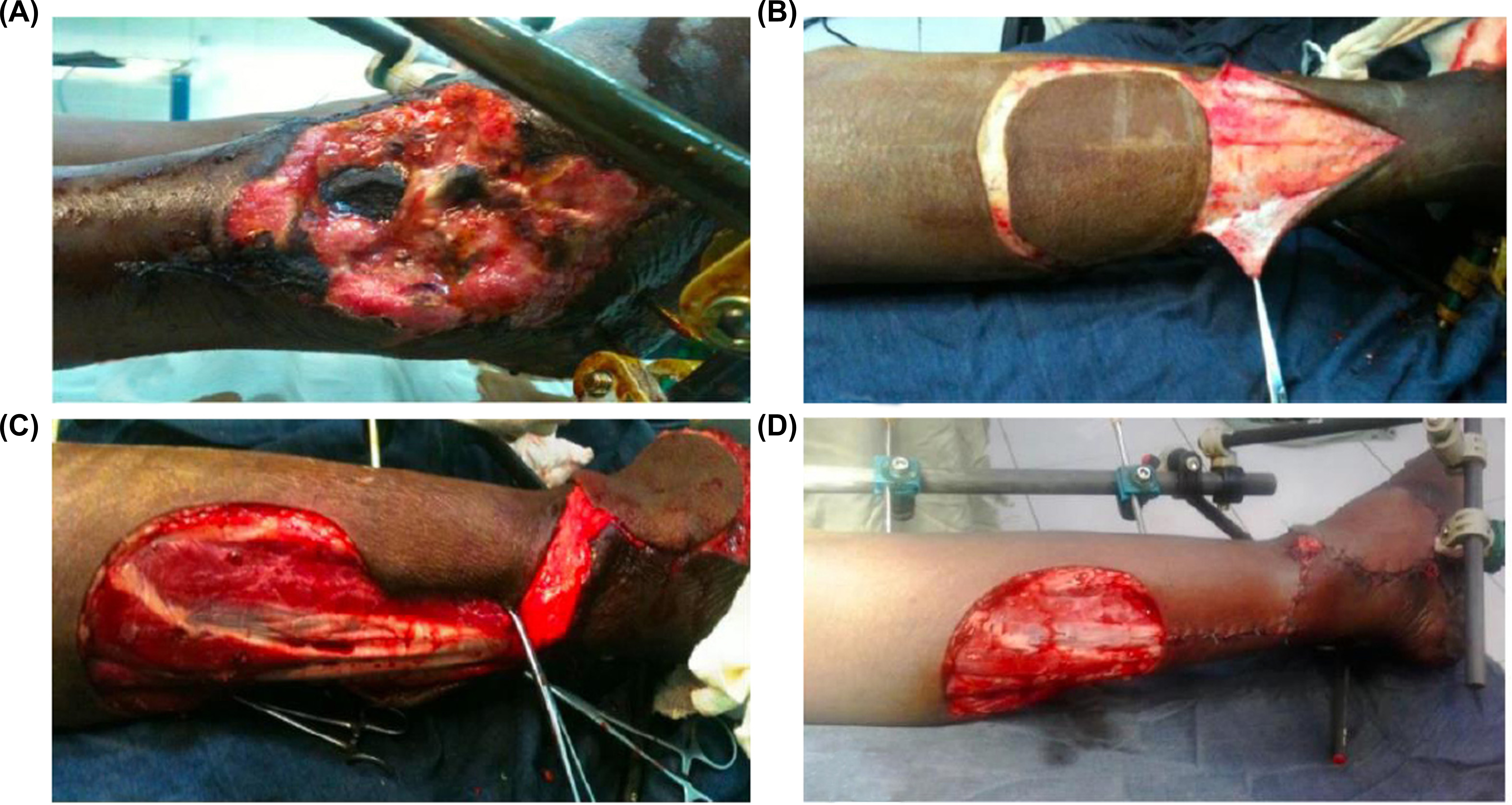
Intraoperative views of the distally based sural flap.
(A) Soft-tissue defect on the lateral aspect of the right ankle.
(B) Sural flap elevation.
(C) placement of the sural flap.
(D) final appearance at the end of the procedure.
Only three flaps (11.5%) were performed during the acute phase, while nine (34.6%) were performed in the subacute phase and fourteen (53.9%) in the chronic phase. Cross-leg (heterolateral) flaps were required in five cases (19.2%).
Regarding the donor site, 24 patients (92.3%) underwent delayed split-thickness skin grafting, while 2 patients (7.7%) achieved directed secondary healing.
Postoperatively, 17 patients (65.4%) achieved uneventful healing with complete flap integration. Venous congestion was the most frequent complication, occurring in 9 patients (34.6%). It progressed to partial necrosis (Fig. 2) in 4 cases (15.4%) and total necrosis in 3 cases (11.5%). Late complications included unsightly scars (3 cases) and ankle stiffness.
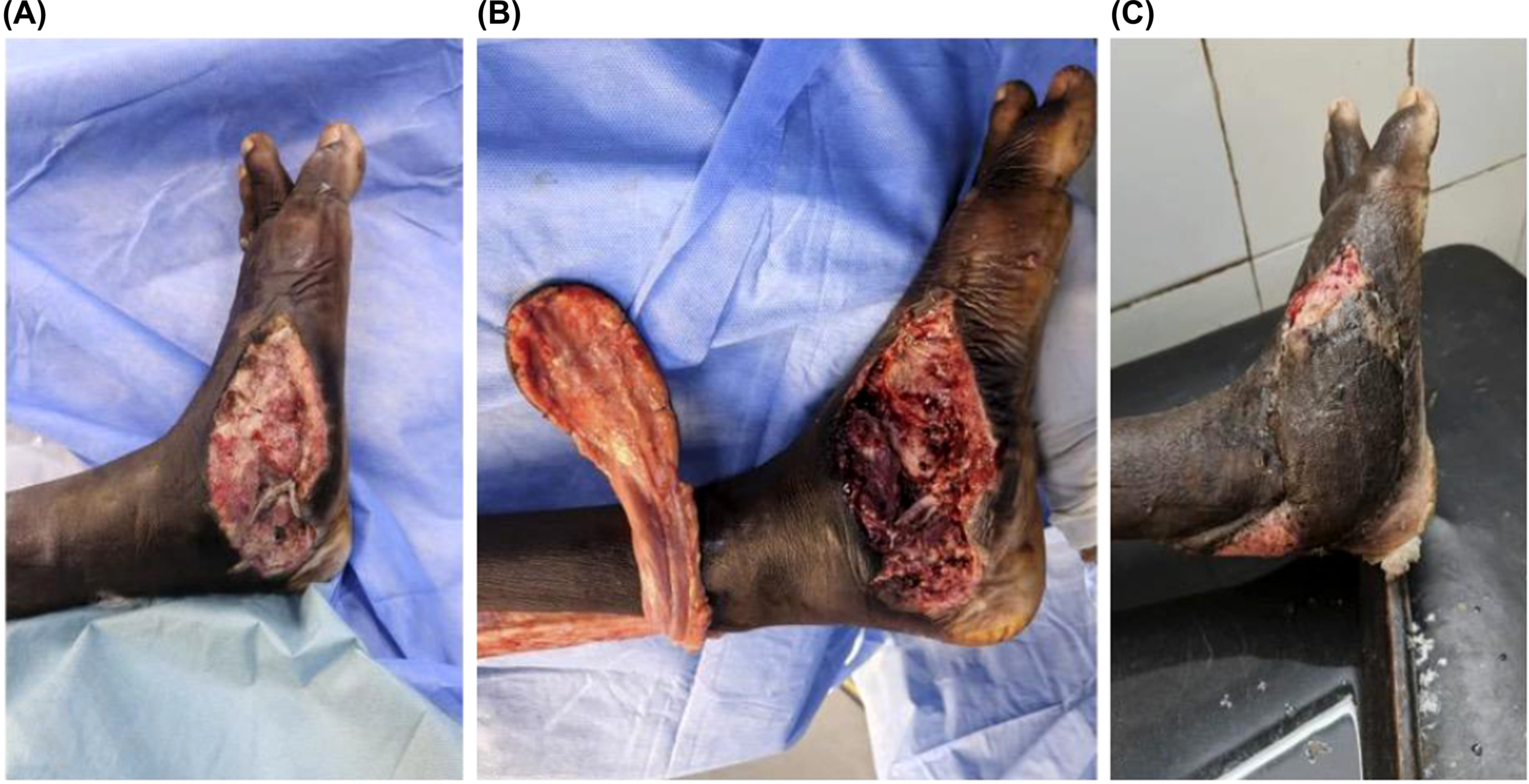
Partial necrosis of the sural flap of the left foot.
(A) Soft-tissue loss of the medial side of the left foot.
(B) Flap elevation and foot debridement.
(C) Sural flap partial necrosis.
Based on the functional and aesthetic evaluation criteria, the mean outcome score was 5.84 ± 1.59 points. The final results were rated as very good in 65.4%, good in 23.1%, and poor in 11.5% of cases, corresponding to a flap survival rate of 88.5%.
No statistically significant association was found between identified risk factors and flap survival (p = 0.069).
4. DISCUSSION
Reconstruction of soft-tissue defects of the distal lower limb remains challenging due to the complex regional anatomy, relatively poor skin vascularity, and limited local muscle coverage [7, 13, 14]. In our series, the sural flap was used mainly to cover defects of the distal third of the leg (50%), followed by the ankle region (42.3%) and the heel (7.7%).
The distal third of the leg and the ankle are commonly reported recipient sites for the sural flap [6, 15, 16]. Daar et al. reported the heel as the most frequent indication, accounting for 40.8% of cases in their systematic review [17]. This discrepancy may reflect differences in case mix, trauma mechanisms, and reconstructive preferences between institutions.
In our study, traumatic soft-tissue loss was the leading indication for sural flap reconstruction. Other reported indications in the literature include postoperative complications, diabetic foot ulcers, tumor resection defects, and burn sequelae [6, 7, 14]. Indeed, trauma remains the most common etiology in most published series [17–20].
The cross-leg flap represents a valuable alternative for complex distal lower-limb reconstruction, particularly when free-flap transfer is contraindicated due to vascular injury or limited microsurgical resources [21]. Within this study population, the technique was deployed in 5 patients (19.2%), representing the definitive, feasible locoregional reconstructive modality available at this institution.
Postoperative complications were dominated by persistent venous congestion (34.6%), followed by partial flap necrosis (15.4%) and total flap necrosis (11.5%). Venous congestion is consistently reported as the most frequent complication of the sural flap. In a recent multicenter study, Abualhaj et al. reported venous congestion in 83.3% of cases, with persistent congestion progressing to partial necrosis in approximately 10% [12]. Similarly, other authors have identified venous congestion and marginal necrosis as the principal complications associated with sural flap reconstruction [6, 8, 15, 22].
In a systematic review including 2,592 sural flaps, Tripathee et al. reported an overall complication rate of 25.2%. Partial flap loss was the most frequent complication (7.85%), followed by venous congestion (3.05%) and complete flap loss (2.5%) [19].
Several patient-related factors have been identified as contributors to venous congestion and flap necrosis, including diabetes mellitus, smoking, peripheral vascular disease, and advanced age. These factors impair microcirculation and venous outflow, thereby increasing the risk of partial or total flap failure [17, 19, 23].
Early interventions such as flap scarification were effective in reducing venous congestion, while local wound care contributed to favorable outcomes in cases of partial necrosis. Total flap necrosis was considered a reconstructive failure and required alternative reconstructive procedures. Several technical modifications have been proposed to enhance flap reliability, including de-epidermization, staged procedures, and preoperative identification of perforating vessels [8, 14, 24].
When compared with large published series, the rates of partial and total necrosis observed in our study remain within the upper range of reported values. The most recent series report partial necrosis rates ranging from 5% to 15% and total flap loss rates between 2% and 8%, highlighting the variability in outcomes depending on patient selection and surgical context [6, 8, 17, 19].
Overall, sural flap reconstruction is associated with high success rates, with reported flap survival exceeding 90% in most series [14, 15, 19, 21, 22, 24]. In this study, the survival rate was slightly lower (88.5%). Similar survival rates have been reported in series dominated by traumatic injuries, absence of preoperative vascular imaging, and delayed reconstruction, supporting the hypothesis that timing of coverage and injury severity play a crucial role in flap outcome [17].
This study has several limitations. Its retrospective design may introduce selection and information bias. The small sample size limits statistical power. Delayed presentation and treatment discontinuation related to traditional practices may have influenced patient selection and outcomes. Furthermore, the involvement of multiple surgeons with varying technical approaches could have affected flap evolution. Finally, unmeasured variables such as flap size, rotation angle, and patient-specific risk factors may have influenced outcomes and the internal validity of the study.
CONCLUSION
The distally based sural flap remains a reliable and versatile option for the reconstruction of soft tissue defects in the distal third of the leg, ankle, and heel. Despite potential complications such as venous congestion or partial necrosis, careful surgical technique, timely coverage, and appropriate postoperative care allow for high rates of flap survival and satisfactory functional and aesthetic outcomes, even in resource-limited settings.
AUTHORS’ CONTRIBUTIONS
The authors confirm their contribution to the paper as follows: L.T., C.O.S.: Study conception and design; I.D., M.B. Data collection; A.B.: Draft manuscript. All authors reviewed the results and approved the final version of the manuscript.
LIST OF ABBREVIATIONS
| RTA | = Road Traffic Accident |
| STL | = Soft-Tissue Loss |
ETHICS APPROVAL AND CONSENT TO PARTICIPATE
Given the non-interventional and observational nature of the study, formal approval from an ethics committee was not required according to local regulations.
CONSENT FOR PUBLICATION
Written informed consent for the use of anonymized clinical data was obtained from all adult patients and from the legal guardians of underage participants at the time of admission, in accordance with institutional policy.
AVAILABILITY OF DATA AND MATERIALS
The data supporting the findings of the article are available from the corresponding author and are not publicly available due to institutional and patient confidentiality restrictions.
ACKNOWLEDGEMENTS
Declared none.