All published articles of this journal are available on ScienceDirect.
Robert Wartenberg Syndrome and Sign: A Review Article
Abstract
Background:
Robert Wartenberg, a European-American neurologist, was born in 1887 and died in 1956. His description of radial sensory nerve compression at the forearm is memorialized as Wartenberg’s syndrome. He recognized that involuntary abduction of the little finger could be caused by ulnar nerve palsy - a finding often called Wartenberg’s sign Syndrome and signs are reviewed, and a brief biography is presented.
Objective:
To review Wartenberg’s sign and Wartenberg’s syndrome.
Discussion:
Compression of the superficial branch of the radial nerve, often called Wartenberg’s syndrome, is characterized by pain, paresthesia, and dysesthesia along the dorsoradial distal forearm. Non-operative treatment can include activity restriction and anti-inflammatory medication. If symptoms persist, surgical decompression of the radial nerve is an option. The abducted posture of the little finger - Wartenberg’s sign - can result from a low ulnar nerve palsy. Tendon transfer can be performed to correct this deformity.
Conclusion:
Compression of the superficial branch of the radial nerve and abducted posture of the little finger were described by Robert Wartenberg and carry his name as eponymous syndrome and sign, respectively.
1. INTRODUCTION
Those who rotate on the hand surgery service at our institution are occasionally quizzed about Wartenberg’s sign and Wartenberg’s syndrome. Some know, and some do not. Here, in one place, we review both for the benefit of medical students, residents (and attending physicians). We also present a brief biography of the man whose name is attached to the syndrome and sign.
2. BIOGRAPHY
Robert Wartenberg was born in 1887 in Grodno (then part of Russia, now part of Belarus) and died in 1956 in San Francisco, California. His death was marked by a number of obituaries that reviewed the remarkable arc of his life [1-3]. He was educated in Europe at universities in Kiel, Munich, Freiberg, and Rostock and graduated from the University of Rostock in 1919. Thereafter, he pursued a career in neurology, ultimately becoming a professor of neurology at the University of Freiberg in 1933 [2, 4]. The obituaries and biographies report that Wartenberg left Europe for the United States in 1935. They note that he was a “refuge from Nazi Germany,” that he “fled” in the face of “rising anti-Semitism,” and that he “escape[d] Nazi Germany” [2-5]. Although not specifically stated in the obituaries at the time of his death and in the subsequent encomiums, one can infer that Wartenberg was Jewish.
From Germany, Wartenberg came to the United States and joined the faculty at the University of California School of Medicine (San Francisco) in 1936, rising to clinical professor of neurology in 1952 [3]. He retired in 1954 and died in 1956 at the age of 69. He was reported as “beloved” by his students and that at medical school, “no lectures were more enthusiastically attended” than his [1, 3].
Few physicians receive glowing obituaries in multiple medical journals as Wartenberg did. Rarer still is the physician whose memory and contributions are recalled in the years and decades after his death. Kuzuhara, in 2014, noted that Wartenberg advocated a minimum of laboratory tests and “best treatment in terms of time, safety, and cost” [5]. This is a timeless advice, regardless of the field of medicine. Maranhao-Filho and Gonik, in 2016, described Wartenberg as an “internationally recognized neurologist” whose academic achievements were “vast” [4]. He was noted to have a “tradition of simplifying the examination” [4]. This is a good advice, regardless of the field of medicine.
J.M.S Pierce in 2018 noted Wartenberg's “international repute” [6]. Burkholder and Boes in 2019 described Wartenberg as a “master clinical neurologist” and noted that a Robert Wartenberg lecture is given annually at the American Academy of Neurology meeting [7].
Robert Wartenberg was a neurologist and an author of more than 150 papers published in German and English. Most do not pertain to hand surgery, but some do, and his contributions to hand surgery are worth recalling.
2.1. Wartenberg’s Syndrome
In 1932 Wartenberg reported 5 cases of mononeuropathy of the superficial branch of the radial nerve [8]. The report was in German. Wartenberg gave it the name “cheiralgia paresthetica” which means hand pain [9, 10].
Ehrilich et al. translated Wartenberg’s paper into English in a condensed form in a 1986 report in the Journal of Hand Surgery [11]. They noted that in the decades subsequent Wartenberg’s paper, there had been a paucity of reports of this condition.
Superficial radial nerve palsy, superficial radial nerve compression, cheiralgia paresthetica are synonymous with what is now often called Wartenberg’s syndrome. Sprofkin used the term “Wartenberg’s disease” [9]. Wartenberg was not the first to describe this problem. Matzdorff reported a case of entrapment of the superficial branch of the radial nerve in 1926. However, Wartenberg’s syndrome is the most commonly used eponym for this problem [12, 13].
The superficial branch of the radial nerve is a purely sensory nerve, a distal branch of the radial nerve.
In the upper arm, the radial nerve traverses behind the humerus, wraps around the humerus laterally, and then crosses anterior to the lateral epicondyle of the humerus in the antecubital fossa region where the nerve divides into the posterior interosseous nerve (motor branch) and superficial branch (sensory). The superficial branch runs superficial to the supinator muscle and then along the ulnar edge and deep to the brachioradialis. At about 9 cm proximal to the radial styloid, the superficial branch of the radial nerve becomes subcutaneous between the brachioradialis and the extensor carpi radial longus tendons. More distally in the forearm, the nerve divides into branches, providing sensation for the dorsoradial aspect of the hand, dorsal aspect at the thumb, index and middle fingers proximal to the distal interphalangeal joints [9, 14-16]. Anthony stated that the superficial branch of the radial nerve is vulnerable to compression anywhere in the forearm [16]. However, where the nerve travels from a deep to superficial location, it is particularly vulnerable to compression from the edge of the brachioradialis [14]. Dellon and Mackinnon stated that the site of entrapment is in the area where the superficial branch of the radial nerve transitions from submuscular to subcutaneous, between the fascia, joining the brachioradialis to the extensor carpi radialis tendon [17].
Anthony et al. stated that compression could occur from fascial bands between the brachioradialis and extensor carpi radialis longus, and from fascial “rings” of the brachioradialis [16].
Patients with Wartenberg’s syndrome typically complain of pain, paresthesia, and dysesthesia along the dorsoradial distal forearm extending onto the dorsal first web space and thumb [9, 14]. Symptoms are often worse with the motion of the wrist [17].
The exact area of symptoms can vary [9]. The overlapping pattern of innervation of the superficial branch of the radial nerve and the lateral antebrachial cutaneous nerve can result in variation in sensory findings [10, 18]. Also, there is variability in the digital innervation of the radial sensory nerve [19]. A positive Tinel sign is frequently elicited with gentle tapping over the superficial branch at the radial nerve in the mid-forearm distal to the brachioradialis muscle belly [17]. Sensory abnormalities are often detected on examination on the dorsoradial aspect of the hand [17]. Forced forearm pronation and forceful hyperpronation of the forearm with ulnar deviation and flexion of the wrist can provoke symptoms [16, 17].
The cause of nerve compression is not always identifiable. Dellon and Mackinnon noted that patients “usually gave a history of either crushing, contusion, or twisting injury to the wrist or forearm or a job-related activity requiring frequent pronation and supination” [17]. External compression from wristwatch [10, 20] and handcuffs [21-23] have been implicated. The clinical course is often self-limiting. Sprofkin described cheiralgia paresthetica as a “benign and ephemeral disorder” [9]. Dang counseled patients, noting that spontaneous resolution was common [14].
In 1954 Sprofkin reported 3 cases with cheiralgia paresthetica (which he called Wartenberg’s disease). All had a sensory deficit in the radial sensory nerve distribution, with varying degrees of pain. No specific treatment was rendered, and all experienced improvement within several weeks [9].
Braidwood, in 1975, reported 12 cases of superficial radial neuropathy, 6 of which resolved without treatment. Two recovered following a cortisone injection. Four were treated by the division of the radial nerve. After transection, patients were reported to have hypesthesia at the base of the thumb (no mention of the loss of sensation on the dorsoradial hand). The author reported that the patients “felt this was a small price to pay for the relief of the burning pain and paresthesia” [24]. Patient acceptance for this outcome is likely to have changed since then.
Dellon and Mackinnon presented a large series - 51 patients (“with 58 entrapments”) who they diagnosed as having entrapment of the radial sensory nerve [17]. Typical complaints included pain, burning, numbness, tingling on the dorsal radial aspect of the hand. On examination, a positive Tinel sign was found over the sensory branch of the radial nerve distal to the brachioradialis muscle belly. The sensation was diminished at the dorsal radial aspect of the hand. Forced pronation of the forearm provoked symptoms. While they reported a positive Finkelstein sign in 96% of hands, only 17% were diagnosed as having first dorsal extensor tenosynovitis. Non-operative treatment included activity restriction and splinting of the wrist and forearm. Some were given anti-inflammatory medication. All their patients were treated non-operatively for at least 6 months. Surgery was performed on 32 patients (35 hands) with good or excellent results in 86%. Surgery was performed via a longitudinal palmar incision, 6 to 8cm in length. The superficial branch of the radial nerve was identified between the brachioradialis and the extensor carpi radialis longus. The fascia joining the brachioradialis to the extensor carpi radialis was completely released, and the nerve was “freed” until it was “loose in the subcutaneous tissue” [17]. None underwent resection of the nerve as an initial form of treatment, although the authors acknowledged that nerve resection might be appropriate in certain circumstances.
Lanzetta and Foucher, in 1993, presented a report of 52 cases (50 patients) of Wartenberg’s syndrome [10]. A diagnosis of deQuervain’s tendonitis was made in 50% of their cases (concomitant diagnoses). The authors of this study felt that Wartenberg’s syndrome occurs “more frequently than might be expected” and that it “is probably too often misdiagnosed as deQuervain’s disease or, in the presence of this condition, it is not diagnosed at all”.
The frequency of Wartenberg’s syndrome is not known. Some feel that isolated neuropathy of the superficial branch of the radial nerve is a rare condition [25]. However, it is not clear whether it is rare or simply rarely diagnosed. Nonetheless, awareness of its existence can facilitate recognition and diagnosis. Initial treatment is often non-operative; surgical decompression of the superficial branch of the radial nerve can result in a favorable outcome.
2.2. Wartenberg Sign
In addition to the eponymous syndrome, Wartenberg’s name is affixed to a sign of ulnar nerve palsy. The sign, as described by Wartenberg, “consists of a position of the abduction ASSUMED by the little finger” [26]. As Wartenberg noted, adduction of the little finger is performed by interosseous muscles, and abduction is performed by hypothenar muscles, both innervated by the ulnar nerve. However, abduction is also performed by the extensor digiti minimi and the extensor digitorum communis to the little finger, both innervated by the radial nerve. If the ulnar nerve is injured, intact radial nerve function can result in unopposed abduction of the little finger [26, 27]. Wartenberg noted that this finding (his sign) is much more pronounced with fingers in extension than flexion [26]. He also noted that occasionally the ring finger could also assume a position of abduction [26].
Akinleye et al. described relative contributions to small finger abduction of ulnar versus radial slip of the extensor digiti minimi [28]. As one might expect, the ulnar slip of the extensor digiti minimi is the major contributor to abduction. Clinically warranted extensor tendon transfer could be performed to correct the abducted posture [29, 30]. It should be noted that not all cases of the abducted little finger are due to ulnar nerve palsy [31]. Alter reported abduction of the little finger in mild hemiparesis and called this the “digiti quinti sign” [32]. Vincent reported a similar finding in patients with hemiplegic migraine [33]. The abducted posture of the little finger can be seen in cases of cervical cord compression [34]. Freeland reported an abducted little finger caused by avulsion of the insertion of the third volar interosseous muscle [35]. These seem to be rarer causes of the abduction of the little finger, and ulnar nerve palsy should be high on the list when evaluating patients with a Wartenberg’s sign.
2.3. Bonus
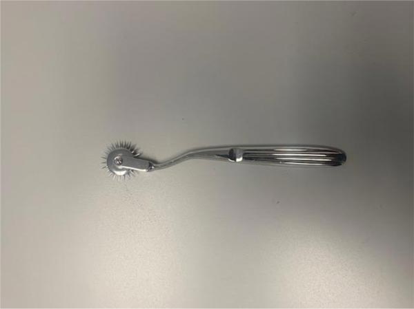
Wartenberg described “a pinwheel, like that used by a tailor in making patterns, useful in the examination for pain sensibility and superficial reflexes” [36]. Sharp pins radiate from the wheel. The wheel rotates as it is rolled across the skin. In examining for pain, the pinwheel is rolled “gently” over the skin. The design of the pinwheel allows skin pricks that are “equal with regard to strength, time and distance”. This device is referred to as a Wartenberg wheel or Wartenberg pinwheel (Fig. 1).

CONCLUSION
Robert Wartenberg was a renowned neurologist whose contribution to orthopedic surgery continues through his eponymous sign and syndrome.
CONSENT FOR PUBLICATION
Not applicable.
FUNDING
None.
CONFLICT OF INTEREST
The authors declare no conflict of interest, financial or otherwise.
ACKNOWLEDGEMENTS
Declared none.


