EDITORIAL


The Acute Occult Salter-Harris Injuries in Children


N. K. Sferopoulos1, *
1 Department of Pediatric Orthopaedics, “G. Gennimatas” Hospital, Thessaloniki, Greece


Article Metrics

CrossRef Citations:
0
Total Statistics:

Full-Text HTML Views: 2861
Abstract HTML Views: 353
PDF Downloads: 882
ePub Downloads: 219
Total Views/Downloads: 4315
Unique Statistics:

Full-Text HTML Views: 1812
Abstract HTML Views: 257
PDF Downloads: 737
ePub Downloads: 177
Total Views/Downloads: 2983


Creative Commons License
© 2019 N. K. Sferopoulos.

open-access license: This is an open access article distributed under the terms of the Creative Commons Attribution 4.0 International Public License (CC-BY 4.0), a copy of which is available at: https://creativecommons.org/licenses/by/4.0/legalcode. This license permits unrestricted use, distribution, and reproduction in any medium, provided the original author and source are credited.

* Address correspondence to this author at the Department of Pediatric Orthopaedics, “G. Gennimatas” Hospital, P. Papageorgiou 3, 546 35, Thessaloniki, Greece; Tel: 00302310963270; Fax: 00302310968265;
E-mail: sferopoulos@yahoo.com


EDITORIAL

Salter and Harris reported their five-part classification in children with open growth plates to grade fractures of the physeal plate according to the involvement of adjacent metaphysis and epiphysis. Their grading is the most commonly used classification for pediatric physeal fractures and is based on radiographic appearance, causal mechanism and prognosis, concerning the disturbance of growth [1-3].

Traditionally, the appearance of a fracture line on plain radiographs is needed for the primary diagnosis of a fracture. In the acute occult or obscure Salter-Harris injuries in children, there is no evidence of a fracture line at the initial anteroposterior and lateral radiographic survey. Occult undisplaced physeal fractures and specific bone bruises are the two subgroups of the acute occult Salter-Harris injuries in children. Such injuries should be clinically suspected, since they are always followed by a varying severity of tenderness, swelling, pain and refusal to use the injured extremity, and appropriately treated [4-10].

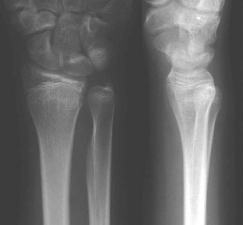
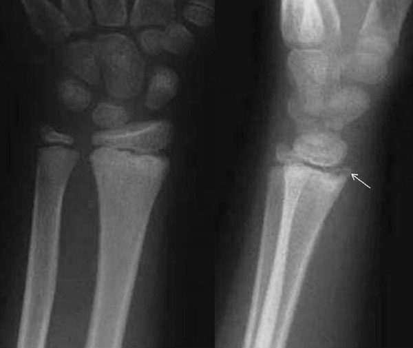
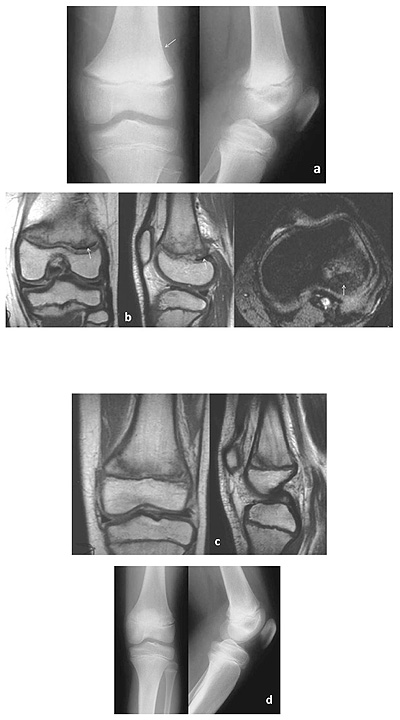
Radiographic diagnosis of the acute occult undisplaced Salter-Harris injuries of the extremities in children is usually secondary, referring to the findings that are consistent with the fracture healing process. These may include signs of periosteal healing and bone sclerosis, as well as the delayed appearance of a fracture line or a widened physeal plate [11-20]. There should be a clear distinction of these findings from the late radiographic signs of physeal injuries that are due to long-term complications, such as altered growth or premature closure of the growth plate, which are usually evident months or even years post-injury. The time of appearance of the radiographic signs of bone healing varies from a few days to 5 weeks, mainly depending on the age of the patient. Periosteal reaction is usually evident at 2 weeks after the injury. The subperiosteal new bone formation is due to the full thickness detachment of the periosteum from the metaphysis and diaphysis (Fig. 1). It is initially separable from the cortex but becomes incorporated in the cortex within a couple of months. Increased density due to the calcific callus formation is usually evident in the metaphyseal segment of the occult physeal injury (Fig. 2). It usually appears 4 to 5 weeks post-injury. The delayed appearance of a fracture line following a physeal injury usually involves the metaphyseal segment of the physeal injury. It is due to reabsorption involving an area of 1 to 2 mm of the bone at the metaphyseal fracture edges that have lost their blood supply. Bone reabsorption makes the metaphyseal fracture line become radiographically distinct, usually 2 to 5 weeks after the injury (Fig. 3). It may also occasionally involve the growth plate, appearing as physeal plate widening (Fig. 1). This radiographic finding should be clearly differentiated from the immediate post-traumatic widening of the physeal plate that is usually due to either a displaced fracture separation (slight widening) or soft tissue interposition (physeal gap of more than 3 mm) [21-23]. Therefore, it should be indicated that the secondary radiographic appearance of the widening of the growth plate may represent a healing sign of an unrecognized physeal plate injury [24, 25]. Physeal widening has also been diagnosed in the knees of high-level child athletes representing a sign of stress injury [26, 27]. It may also occasionally represent a significant predictor of subsequent growth disturbance [28].

Careful patient examination is particularly important in the acute occult undisplaced Salter-Harris injuries. Supplemental imaging, which usually includes oblique (tangential) projections, computerized tomography (CT) and magnetic resonance imaging (MRI), has been used to determine the precise fracture pattern in these injuries [29-32]. The detection of a fracture line on supplemental imaging indicates a subtle fracture. However, supplemental imaging rarely adds information that influences the treatment strategy of these fractures. Therefore, it is our standard policy not to perform other imaging studies, than the initial radiographic evaluation, in order to minimize the radiation dose and the imaging costs, and to treat all the injuries in children, in whom a potential fracture is clinically, but not radiographically, diagnosed with the appropriate immobilization.

The Salter and Harris classification scheme includes two acute injuries that may remain occult on conventional radiography in both the initial and follow-up evaluation. They are the undisplaced type I and the type V lesions.

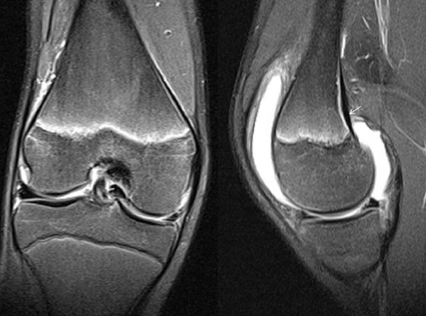
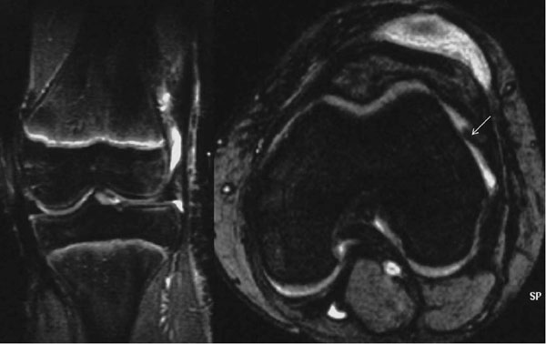
The Salter-Harris type I lesion represents a complete or incomplete fracture through the physeal plate without any bone fracture. It implies a shearing injury, although an avulsion or rotational injury cannot be excluded [1, 33]. When type I physeal injury occurs and the applied load is sufficient to disrupt the epiphyseal-metaphyseal unit, the periosteum may strip from the underlying bony cortex. This represents the earliest stages of physeal failure, preceding the failure of the continuity of perichondrium with the periosteum and displacement [5]. Radiographs are negative at the time of assessment in undisplaced injuries except for an associated soft tissue swelling. Stress radiographs to document displacement are generally unnecessary and probably unwise. The value of MRI was evaluated in a 9-year-old girl with a potential type I injury of the distal femur. The findings included widening of the physeal plate, perichondrial disruption, intracartilaginous fracture, and adjacent metaphyseal edema indicating a Salter-Harris type I fracture without displacement [34]. Other reports have described the MRI findings following soft tissue entrapment in displaced Salter-Harris type I injuries [35, 36]. It may be reasonable to consider that the nature of an undisplaced complete type I injury is most likely to be an initially minimally displaced lesion, in which the continuity of the perichondrium with the periosteum is preserved and the reduction is restored due to the elasticity of the perichondrium-periosteum complex. Therefore, it is mandatory to consider the MRI diagnosis of subperiosteal haematoma due to an even minor local metaphyseal detachment of the periosteum from the metaphyseal bone, as an initial absolute prerequisite for the diagnosis of an undisplaced complete type I growth plate injury (Fig. 4). Furthermore, it might be prudent to consider that a local minor disruption of the periosteum on the MRI findings can also be consistent with an undisplaced complete type I growth plate injury (Fig. 5). On the other hand, Salter-Harris type I incomplete physeal fractures may be associated with a pathological signal of the disrupted portion of the physeal plate but not with subperiosteal haematoma or a disrupted perichondrium-periosteum unit.

The pathology of an undisplaced Salter-Harris type II injury can be similar to that of a type I injury (Fig. 6). The roughly triangular variably-sized Thurston-Holland metaphyseal fragment is recognized as the hallmark of the Salter-Harris II injury [1, 37], while children with occult SH type II fractures of the distal femur that were evident on the MRI examination were all found to have posterior periosteal disruption and other soft tissue findings, thus indicating a hyperextension mechanism of injury [38].

Fig. (1). A 10-year-old boy forced his wrist into full extension while playing football. Clinical examination was indicative of a physeal fracture of the distal radius. The radiographs were negative. He was treated with a splint. Radiographs 5 weeks post-injury indicated a Salter-Harris type II lesion. The findings included widening of the distal radial physis on both views, widening between the palmar metaphyseal flake fragment and the metaphysis on the lateral view, and periosteal reaction on the palmar aspect of the radius (arrow).

Fig. (2). A 14-year-old girl stressed her wrist against a wall while running. Clinical examination was indicative of a physeal fracture of the distal radius. Radiographs were negative. She was treated with a splint. Radiographs 5 weeks post-injury were indicative of distal radial metaphyseal sclerosis, most likely due to an occult Salter-Harris type II injury of the distal radius.

Fig. (3). A 7-year-old girl entrapped her wrist in a swing. Clinical examination was indicative of a physeal fracture of the distal radius. Radiographs were negative. She was treated with a splint. The radiographs 5 weeks post-injury indicated a Salter-Harris type II lesion. The findings included widening between the palmar metaphyseal flake fragment and the metaphysis (arrow), as well as periosteal reaction on the palmar aspect of the distal radius.

Fig. (4). A 14-year-old boy sustained a rotational injury of the knee while playing football. Clinical examination was indicative of a physeal fracture of the distal femur. Radiographs were negative. A MRI was performed 6 weeks post-injury. The fat-suppressed images indicated knee effusion, widening and irregularity of the distal femoral growth plate demonstrating edema (high signal) along the physis, which indicates injury (coronal view) and uplifting of the periosteum from the posterior surface of the medial femoral condyle (arrow) with a small subperiosteal collection (sagittal view), most likely due to an occult Salter-Harris type I injury of the distal femur. No disruption and irregularity of the periosteum-perichondrium complex was noted.

Fig. (5). A 12-year-old girl fell while running with a bicycle. Clinical examination was indicative of a physeal fracture of the distal femur. Radiographs were negative. A MRI was performed 6 weeks post-injury. The fat-suppressed images indicated a prepatellar traumatic subcutaneous hematoma, widening and irregularity of the distal femoral growth plate demonstrating edema (high signal) along the physis, which indicates injury (coronal view) and local disruption of the periosteum (arrow) of the lateral femoral condyle (axial view), most likely due to an occult Salter-Harris type I injury of the distal femur.

Fig. (6). An 11-year-old boy injured his knee after a fall while playing football. Initial radiographs indicated no evidence of a fracture line. A clinical diagnosis of a potential Salter-Harris type I injury was made. Radiographs, 5 weeks post-injury, revealed widening of the distal femoral growth plate and periosteal reaction (arrow) along the distal lateral femoral metaphysis (a). A MRI was performed 6 weeks post-injury. Proton density (coronal and sagittal), as well as FL2D (axial) views, indicated a hypointense membrane interposed within the posterolateral portion of the disrupted physis, consistent with entrapped periosteum (arrows). There was bone bruising of the lateral femoral condyle (b). Proton density (coronal and sagittal) images also confirmed a medial metaphyseal Thurston-Holland fracture fragment with adjacent periosteal elevation (c). Radiographs, 2 years post-injury, showed incorporation of the periosteal reaction in the cortex and no altered growth (d). The final diagnosis was a Salter-Harris type II injury of the distal femur.

Fig. (7). A 13-year-old boy injured his wrist after a fall while playing football. Plain radiographs at injury were negative (a). Radiographs following 5 weeks of immobilization indicated metaphyseal sclerosis in close proximity to the distal radial physis (b). CT findings at 5 weeks after injury were indicative of a healing undisplaced Salter-Harris type II fracture of the distal radius with a flake palmar metaphyseal fragment (c).

Compared with radiography and CT, MRI is more expensive, but it is recommended in the literature because it provides a noninvasive means for the detection of the Salter-Harris acute occult fractures and may also alter the Salter-Harris staging [6, 29-31].

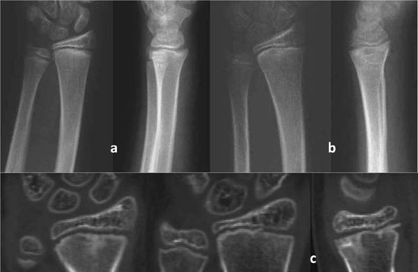
In addition, the diagnostic criteria were evaluated in the patients who were initially diagnosed with a complete undisplaced Salter-Harris type I lesion from the hospital database. In most of the cases, the diagnosis was based on the clinical findings. A small number of these patients were evaluated with a CT. In all of them, the diagnosis was changed to a Salter-Harris type II lesion due to the detection of a tiny or flake metaphyseal fragment that was attached to the epiphysis (Fig. 7). It was realized that the value of MRI to define a metaphyseal fragment becomes less with tiny or flake metaphyseal fragments. On the other hand, the value of CT to visualize tiny metaphyseal fragments is not limited. Therefore, it may be prudent to consider all the radiographically-diagnosed complete undisplaced Salter-Harris type I lesions as potential type II injuries until a CT scan proves the contrary, whenever the CT examination value is considered sufficiently reasonable.

The Salter-Harris type V lesion represents a radiographically occult compression injury of the growth plate, following considerable axial load [39]. This should be differentiated from the crushing of physeal cells that may occur in any other type of Salter-Harris physeal fracture [40-47]. The definition that the type V injury is a longitudinal compressive force on the physis damaging only the physeal cells, but not a fracture since no trabeculae are broken [48, 49] has been questioned. This statement was not consistent with the description of type V injury given by Salter and Harris in their original article, who defined it as an injury resulting from a very severe crushing force applied through the epiphysis to an area of the epiphyseal plate [1]. The first inclusion criterion to diagnose a type V lesion, requiring normal findings on roentgenograms taken in at least two planes at the time of injury [49], should also be reevaluated. It would probably be prudent to change it to indicate no evidence of a fracture line to the physis, epiphysis or metaphysis. The two other inclusion criteria indicating that the patient has not received any form of manual treatment and that a retrospective diagnosis is made only after premature partial or complete growth arrest, secondary to physeal bridge formation, are absolute prerequisites to define a type V injury. The radiographic and/or CT appearance of impaction or diminution of the width of the physis associated with no evidence of a fracture line, as well as the persistence of significant pain at the level of the physeal plate following a period of immobilization, exceeding 5 to 6 weeks, for an occult physeal injury could be absolute indications to perform a MRI. The appearance of two significant bone contusions opposing one another on either side of the injured physeal plate, associated with local diminished width of the growth plate, has been considered to be pathognomonic of a Salter-Harris type V injury [50, 51]. The primary etiological factor of a type V lesion has been supposed to be ischemic damage rather than direct cellular compression in

Fig. (8). A 13-year-old girl injured her knee after a fall while playing volleyball. Radiographs at injury were negative. The high severity of the clinical symptoms and signs 5 weeks post-injury necessitated a MRI examination. The fat-suppressed images revealed two circumscribed bone contusions opposing one another on either side of the medial part of the proximal tibial growth plate (coronal view) and local diminished width of the proximal tibial growth plate (sagittal view). The findings were considered pathognomonic of a type V injury (a). A CT scan performed a year post-injury indicated a physeal bar at the site of the injury, as well as fusing of the central part of the proximal tibial growth plate (b). No clinical or radiographic abnormality was detected after complete closure of the proximal tibial growth plate.

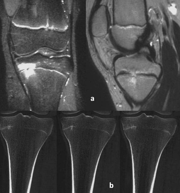
the germinal zone [49]. Histologic findings of the distal radius and ulna supported this concept suggesting that microscopic trauma may lead to microbridges in the form of transphyseal linear ossific striations, which may not necessarily lead to a complete arrest. However, they may interfere with growth leading to the well-recognized variations in the relative length of the distal radius and ulna [52]. In addition, growth disturbance is not encountered whenever the Salter-Harris type V injury occurs at the end of the growth (Fig. 8).

With the advent of fat-suppression techniques, a new category of injury has also been recognized on MRI. It refers to bone bruise or contusion, which usually involves a well-circumscribed area of cancellous bone. The nature of the high intra-osseous signal intensity on MRI using fat suppression is likely to represent focal edema and hemorrhage, following a microscopic trabecular injury and hemorrhage of bone marrow without disruption of adjacent cortices or articular cartilage [53-57].

The value of MRI was evaluated to diagnose non-radiographically evident injuries of the distal forearm and wrist, as well as of the ankle and foot, in children whose symptoms did not resolve after 5 to 6 weeks of immobilization. The MRI examination indicated the occult lesion of bone bruising in all the patients. It was suggested that the Salter-Harris type I complete undisplaced injuries were consistent with the MRI detection of bone bruising involving the metaphysis in close proximity to the distal radial growth plate, while bone bruises localized on both sides of the growth plate were considered as potential Salter-Harris type V injuries [58-60].

Finally, it may be prudent to extend the findings of this report to all the classification schemes for physeal injuries in children that have been reported in the literature, either before or after the Salter-Harris classification [61-71]. Our recommended mode of treatment for pediatric physeal injuries of the extremities when a fracture is clinically, but not radiographically, evident is splint immobilization to prevent displacement and allow fracture healing. The patient is reexamined clinically and radiologically once a week for a two-week period. In the cases that radiographic findings of bone healing are evident, as well as patients with no radiographic findings in the follow-up evaluation, but with persistent clinical symptoms, the immobilization period is prolonged for a sufficient period. In the latter, the period to resolve the clinical symptoms depends on the patient’s age. The period that is usually required for the clinical symptoms and signs to resolve following an acute occult physeal injury, which is more likely to be a Salter-Harris type II injury, varies between 4 weeks for the preschool ages to 5 weeks for older children. Children with persistent symptoms following a period of immobilization that would be sufficient for the healing of an acute physeal injury may suffer from bone bruising. The review of the related literature indicates that a limited MRI examination is useful and valuable in the detection of bone bruising in children with persistent symptoms following 5 to 6 weeks of immobilization.

CONCLUSION

In conclusion, the most challenging aspect of MRI-based diagnosis of the acute occult physeal injuries is the recognition of rare undisplaced complete Salter-Harris type I and type V injuries, as well as of bone bruising.

Traditionally, the appearance of a fracture line or signs of fracture healing on plain radiographs was needed to diagnose a fracture. Thus, children suffering from bone contusions might have been encouraged to use an injured extremity or to participate in sporting activities after an insufficient period of immobilization. An elongated immobilization and recovery period is the only required mode of treatment until complete resolution of the clinical symptoms and signs to prevent the subsequent development of insufficiency fractures. Bone bruising represents a distinct clinical entity with ‘benign’ characteristics since no long-term morbidity occurred in any of the reported patients. The single exception to this rule is the bone bruises which are associated with a Salter-Harris type V physeal injury in the growing skeleton.

CONFLICT OF INTEREST

The author certifies that he has no commercial associations (such as consultancies, stock ownership, equity interest, patent/licensing arrangements, etc.) that might pose a conflict of interest in connection with the submitted article. The author received no financial support for this study.

ACKNOWLEDGEMENTS

Declared none.

REFERENCES

[1] Salter RB, Harris WR. Injuries involving the epiphyseal plate. J Bone Joint Surg Am 1963; 45(3): 587-622.
[2] Rogers LF. The radiography of epiphyseal injuries. Radiology 1970; 96(2): 289-99.
[3] Rogers LF, Poznanski AK. Imaging of epiphyseal injuries. Radiology 1994; 191(2): 297-308.
[4] Simpson WC Jr, Fardon DF. Obscure distal femoral epiphyseal injury. South Med J 1976; 69(10): 1338-40.
[5] Ogden JA, Ganey T, Light TR, Southwick WO. The pathology of acute chondro-osseous injury in the child. Yale J Biol Med 1993; 66(3): 219-33.
[6] Naranja RJ Jr, Gregg JR, Dormans JP, Drummond DS, Davidson RS, Hahn M. Pediatric fracture without radiographic abnormality. Description and significance. Clin Orthop Relat Res 1997; (342): 141-6.
[7] Ogden JA. Skeletal injury in the child 2000.
[8] Boutis K, Komar L, Jaramillo D, et al. Sensitivity of a clinical examination to predict need for radiography in children with ankle injuries: a prospective study. Lancet 2001; 358(9299): 2118-21.
[9] Herring A. Tachdjian’s pediatric orthopaedics 3rd ed. 2002.
[10] Elvey M, Patel S, Avisar E, White WJ, Sorene E. Defining occult injuries of the distal forearm and wrist in children. J Child Orthop 2016; 10(3): 227-33.
[11] Charnley J. The closed treatment of common fractures 1950.
[12] Trueta J. Studies of the development and decay of the human frame 1968.
[13] Islam O, Soboleski D, Symons S, Davidson LK, Ashworth MA, Babyn P. Development and duration of radiographic signs of bone healing in children. AJR Am J Roentgenol 2000; 175(1): 75-8.
[14] Wilkins KE. Principles of fracture remodeling in children. Injury 2005; 36(Suppl. 1): A3-11.
[15] Staheli LT. Practice of pediatric orthopedics 2nd ed. 2006.
[16] Augustin G, Antabak A, Davila S. The periosteum. Part 1: Anatomy, histology and molecular biology. Injury 2007; 38(10): 1115-30.
[17] Kleinman PK. Problems in the diagnosis of metaphyseal fractures. Pediatr Radiol 2008; 38(Suppl. 3): S388-94.
[18] Dwek JR. The periosteum: What is it, where is it, and what mimics it in its absence? Skeletal Radiol 2010; 39(4): 319-23.
[19] Halliday KE, Broderick NJ, Somers JM, Hawkes R. Dating fractures in infants. Clin Radiol 2011; 66(11): 1049-54.
[20] Prosser I, Lawson Z, Evans A, et al. A timetable for the radiologic features of fracture healing in young children. AJR Am J Roentgenol 2012; 198(5): 1014-20.
[21] McAnally JL, Eberhardt SC, Mlady GW, Fitzpatrick J, Bosch P. Medial collateral ligament tear entrapped within a proximal tibial physeal separation: Imaging findings and operative reduction. Skeletal Radiol 2008; 37(10): 943-6.
[22] Segal LS, Shrader MW. Periosteal entrapment in distal femoral physeal fractures: harbinger for premature physeal arrest? Acta Orthop Belg 2011; 77(5): 684-90.
[23] Li X, Aubin ME, Curry EJ. Mortimer, Errol SM. Entrapped periosteum preventing reduction of a Salter-Harris II distal tibial fracture in an adolescent patient. MRI and intra-operative findings. Curr Orthop Pract 2011; 22(6): 582-6.
[24] Laor T, Hartman AL, Jaramillo D. Local physeal widening on MR imaging: an incidental finding suggesting prior metaphyseal insult. Pediatr Radiol 1997; 27(8): 654-62.
[25] Laor T, Jaramillo D. MR imaging insights into skeletal maturation: what is normal? Radiology 2009; 250(1): 28-38.
[26] Laor T, Wall EJ, Vu LP. Physeal widening in the knee due to stress injury in child athletes. AJR Am J Roentgenol 2006; 186(5): 1260-4.
[27] Jawetz ST, Shah PH, Potter HG. Imaging of physeal injury: overuse. Sports Health 2015; 7(2): 142-53.
[28] Sferopoulos NK. Concomitant physeal fractures of the distal femur and proximal tibia. Skeletal Radiol 2005; 34(7): 427-30.
[29] Jaramillo D, Hoffer FA, Shapiro F, Rand F. MR imaging of fractures of the growth plate. AJR Am J Roentgenol 1990; 155(6): 1261-5.
[30] Carey J, Spence L, Blickman H, Eustace S. MRI of pediatric growth plate injury: correlation with plain film radiographs and clinical outcome. Skeletal Radiol 1998; 27(5): 250-5.
[31] Jaramillo D, Kammen BF, Shapiro F. Cartilaginous path of physeal fracture-separations: evaluation with MR imaging-an experimental study with histologic correlation in rabbits. Radiology 2000; 215(2): 504-11.
[32] Close BJ, Strouse PJ. MR of physeal fractures of the adolescent knee. Pediatr Radiol 2000; 30(11): 756-62.
[33] Blagg SE, Giddins GE. Rotational Salter-Harris type 1 fracture of the proximal phalanx. J Hand Surg [Br] 1998; 23(6): 806-7.
[34] Chauvin N, Jaramillo D. Occult distal femoral physeal injury with disruption of the perichondrium. J Comput Assist Tomogr 2012; 36(3): 310-2.
[35] Whan A, Breidahl W, Janes G. MRI of trapped periosteum in a proximal tibial physeal injury of a pediatric patient. AJR Am J Roentgenol 2003; 181(5): 1397-9.
[36] Moura MVT. Trapped periosteum in a distal femoral physeal injury: magnetic resonance imaging evaluation. Radiol Bras 2012; 45(3): 184-6.
[37] Ogden JA. Injury to the growth mechanisms of the immature skeleton. Skeletal Radiol 1981; 6(4): 237-53.
[38] Kritsaneepaiboon S, Shah R, Murray MM, Kleinman PK. Posterior periosteal disruption in Salter-Harris Type II fractures of the distal femur: evidence for a hyperextension mechanism. AJR Am J Roentgenol 2009; 193(6): W540-5.
[39] Letts RM. Compression injuries of the growth plate. Behavior of the growth plate 1988.
[40] Peterson HA. Premature closure of the distal radial physis after fracture of the distal radial metaphysis. J Pediatr Orthop 1997; 17(1): 126-7.
[41] Ecklund K, Jaramillo D. Imaging of growth disturbance in children. Radiol Clin North Am 2001; 39(4): 823-41.
[42] Cannata G, De Maio F, Mancini F, Ippolito E. Physeal fractures of the distal radius and ulna: long-term prognosis. J Orthop Trauma 2003; 17(3): 172-9.
[43] Lee TM, Mehlman CT. Hyphenated history: Park-Harris growth arrest lines. Am J Orthop 2003; 32(8): 408-11.
[44] Sailhan F, Chotel F, Guibal AL, et al. Three-dimensional MR imaging in the assessment of physeal growth arrest. Eur Radiol 2004; 14(9): 1600-8.
[45] Basener CJ, Mehlman CT, DiPasquale TG. Growth disturbance after distal femoral growth plate fractures in children: a meta-analysis. J Orthop Trauma 2009; 23(9): 663-7.
[46] Rathjen KE, Birch JG. Physeal injuries and growth disturbances. Rockwood and Wilkins’ Fractures in children 7th ed. 2010.
[47] Kennon JC, Ganey TM, Gaston RG, Ogden JA. Continued growth after limited physeal bridging. J Pediatr Orthop 2013; 33(8): 857-61.
[48] Peterson HA. Physeal fractures: Part 3. Classification. J Pediatr Orthop 1994; 14(4): 439-48.
[49] Peterson HA, Burkhart SS. Compression injury of the epiphyseal growth plate: fact or fiction? J Pediatr Orthop 1981; 1(4): 377-84.
[50] Sferopoulos NK. Type V physeal injury. J Trauma 2007; 63(6): E121-3.
[51] Sferopoulos NK. The classification of physeal injuries. ARC Journal of Orthopedics 2016; 1(1): 23-7.
[52] Ogden JA. Transphyseal linear ossific striations of the distal radius and ulna. Skeletal Radiol 1990; 19(3): 173-80.
[53] Meyers SP, Wiener SN. Magnetic resonance imaging features of fractures using the short tau inversion recovery (STIR) sequence: correlation with radiographic findings. Skeletal Radiol 1991; 20(7): 499-507.
[54] Nakamae A, Engebretsen L, Bahr R, Krosshaug T, Ochi M. Natural history of bone bruises after acute knee injury: Clinical outcome and histopathological findings. Knee Surg Sports Traumatol Arthrosc 2006; 14(12): 1252-8.
[55] Boutis K, Narayanan UG, Dong FF, et al. Magnetic resonance imaging of clinically suspected Salter-Harris I fracture of the distal fibula. Injury 2010; 41(8): 852-6.
[56] Little JT, Klionsky NB, Chaturvedi A, Soral A, Chaturvedi A. Pediatric distal forearm and wrist injury: An imaging review. Radiographics 2014; 34(2): 472-90.
[57] Lurie B, Koff MF, Shah P, et al. Three-dimensional magnetic resonance imaging of physeal injury: Reliability and clinical utility. J Pediatr Orthop 2014; 34(3): 239-45.
[58] Sferopoulos NK. Bone bruising of the distal forearm and wrist in children. Injury 2009; 40(6): 631-7.
[59] Sferopoulos NK. Residual symptoms of occult pediatric ankle injuries: MRI-diagnosed bone bruising. Br J Med Med Res 2014; 4(1): 295-303.
[60] Sferopoulos NK. Skin burns following cryotherapy in misdiagnosed pediatric injuries. J Bodyw Mov Ther 2018; 22(3): 556-9.
[61] Rang M. The growth plate and its disorders 1968.
[62] Weber BG. Treatment of fractures in children and adolescents 1980.
[63] Shapiro F. Epiphyseal growth plate fracture-separations: A pathophysiologic approach. Orthopedics 1982; 5(6): 720-36.
[64] Rang M. Injuries of the epiphyses, the growth plate, and the perichondral ring. Children’s fractures 2nd ed. 1983.
[65] Peterson HA. Physeal fractures: Part 2. Two previously unclassified types. J Pediatr Orthop 1994; 14(4): 431-8.
[66] Canale ST. Physeal injuries. Green NE, Swiontkowski MF, eds Skeletal trauma in children 3rd ed. 2003.
[67] Rang M, Pring ME, Wenger DR. Rang’s children’s fractures 2005.
[68] Peterson HA. Epiphyseal growth plate fractures 2007.
[69] Sferopoulos NK. Classification of distal radius physeal fractures not included in the Salter-Harris system. Open Orthop J 2014; 8: 219-24.
[70] Cepela DJ, Tartaglione JP, Dooley TP, Patel PN. Classifications in brief: Salter-Harris classification of pediatric physeal fractures. Clin Orthop Relat Res 2016; 474(11): 2531-7.
[71] Levine RH, Foris LA, Waseem M. Fracture, Salter Harris StatPearls 2019.