All published articles of this journal are available on ScienceDirect.
Effect of Osteonecrosis Intervention Rod Versus Core Decompression Using Multiple Small Drill Holes on Early Stages of Necrosis of the Femoral Head: A Prospective Study on a Series of 60 Patients with a Minimum 1-Year-Follow-Up
Abstract
Introduction:
The conventional CD used 10 mm drill holes associated with a lack of structural support. Thus, alternative methods such as a tantalum implant, small drill holes, and biological treatment were developed to prevent deterioration of the joint. The treatment of CD by multiple 3.2 mm drill holes could reduce the femoral neck fracture and partial weight bearing was allowed. This study was aimed to evaluate the effect of osteonecrosis intervention rod versus core decompression using multiple small drill holes on early stages of necrosis of the femoral head.
Method:
From January 2011 to January 2012, 60 patients undergoing surgery for osteonecrosis with core decompression were randomly assigned into 2 groups based on the type of core decompression used: (1) a total of 30 osteonecrosis patients (with 16 hips on Steinburg stageⅠ,20 hips on Steinburg stageⅡ) were treated with a porous tantalum rod insertion. The diameter of the drill hole for the intervention rod was 10mm.(2) a total of 30 osteonecrosis patients (with 14 hips on Steinburg stageⅠ,20 hips on Steinburg stageⅡ) were treated with core decompression using five drill holes on the lateral femur, the diameter of the hole was 3.2 mm. The average age of the patient was 32.6 years (20-45 years) and the average time of follow-up was 25.6 months (12- 28 months) in the rod implanted group. The average age of the patient was 35.2 years (22- 43 years) and the average time of follow-up was 26.3 months (12-28 months) in the small drill holes group.
Results:
The average of surgical time was 40 min, and the mean volume of blood loss was 30 ml in both surgical groups. The average of Harris score was improved from 56.2 ± 7.1 preoperative to 80.2 ± 11.4 at the last follow-up in the rod implanted group (p < 0.05). The mean Harris score was improved from 53.8 ± 6.6 preoperative to 79.7 ± 13.2 at the last follow-up in the small drill holes group (p<0. 05). No significant difference was observed in Harris score between the two groups. At the last follow-up, 28 of 36 hips were at the same radiographic stages as pre-operation, and 8 deteriorated in the rod implanted group. 26 of 34 hips were at the same radiographic stage as pre-operation, and 8 deteriorated in the small drill holes group. No significant difference was observed in radiographic stage between the two groups. There was no favourable result on the outcome of a tantalum intervention implant compared to multiple small drill holes.
Discussion:
CD via multiple small drill holes would allow similar postoperative load-bearing and seems to result in similar or even better clinical outcome without the prolonged implantation of an expensive tantalum implant. A tantalum rod intervention and core decompression using multiple small drill holes were effective on the stage I hips rather than stage II hips.
INTRODUCTION
Using core decompression (CD) for the treatment of early stages of necrosis of the femoral head was an effective method developed by Ficat and Arlet in 1962. CD could reduce the pressure of the femoral head and contribute to the blood reperfusion [1]. However, there were not enough promising results confirmed by other studies. The conventional CD used 10 mm drill holes associated with a lack of structural support. Thus, alternative methods, such asa tantalum implant, small drill holes, and biological treatment were developed to prevent deterioration of the joint [2]. The treatment of CD by multiple 3.2 mm drill holes could reduce the femoral neck fracture and partial weight bearing was allowed [3]. Therefore, we evaluate the effect of osteonecrosis intervention rod versus core decompression using multiple small drill holes on early stages of necrosis of the femoral head.
METHODS
Characteristics of Patients
This prospective study was conducted from January 2011 to January 2012. Inclusion criteria were defined as patients with femoral head osteonecrosis with no evidence of femoral head collapse. The included patients with “ONFH” were chosen by radiological and clinical evidence. MRIs were available for all patients; the Steinburg classification system was used. Patients with stage III and IV were excluded [4] and an age above 65 years.
Therefore, a total of 60 patients undergoing surgery for osteonecrosis with core decompression were randomly classified into 2 groups based on the type of core decompression used: (1) a total of 30 osteonecrosis patients (with 16 hips on Steinburg stage, 20 hips on Steinburg stageⅡ) were treated with a porous tantalum rod insertion. (2) a total of 30 osteonecrosis patients (with 14 hips on Steinburg stage Ⅰ,20 hips on Steinburg stageⅡ) were treated with core decompression using five drill holes on the lateral femur, the diameter of the hole was 3.2 mm. The average age was 32.6 years (20-45 years) and the average follow-up time was 25.6 months (12- 28 months) in the rod implanted group. The average age was 35.2 years (22-43 years) and the average follow-up time was 26.3 months (12-28 months) in the small drill holes group. There were 25 male and 35 female study participants, the male:female ratio was similar for both groups. This study included the following patients, according to the Steinburg Classification System. The preoperative Steinberg stages were as follows: stage I in 30 hips, stage II in 40 hips, and stage III in 0 hips. Osteonecrosis was idiopathic in 15 hips, secondary to steroid use in 46 hips, and associated with alcohol use in 9 hips (Table 1).
Characteristics of patients.
| The Rod Implanted Group | Small Drill Holes Group | p Value | |
|---|---|---|---|
| Number of patients | 30 | 30 | |
| Sex | |||
| Male | 12 | 13 | 0.24 |
| Female | 18 | 17 | |
| Age, mean, years | 32.6±6.3 | 35.2±5.8 | 0.17 |
| Symptom duration, mean, months | 14.2±3.5 | 15.8±4.7 | 0.22 |
| Harris score before surgery | 56.2 ± 7.1 | 53.8±6.6 | 0.31 |
| Harris score after surgery (last follow-up) | 80.2 ± 11.4 | 79.7 ± 13.2 | 0.38 |
| Stage I | 16 | 20 | 0.45 |
| Stage II | 14 | 20 | |
| Etiolgy alcohol | 4 | 5 | 0.25 |
| Etiolgy idiopathic | 7 | 8 | |
| Etiolgy steroid | 22 | 24 | |
| Follow-up after surgery, mean, months | 19.8±4.1 | 18.1±5.2 | 0.32 |
Harris score and survival time.
| Group | Harris Score Improvement | pValue | Survival Rate | Survival Time (M) | pValue |
|---|---|---|---|---|---|
| The rod implanted group | 24.0 ± 7.3 | >0.05 | 28/36(77.8%) | 22.9 | >0.05 |
| Small drill holes group | 25.9 ± 6.6 | 26/34(76.5%) | 22.5 | ||
| Stage I | 30.4±7.7 | <0.05 | 28/30(93.3%) | 25.1 | <0.05 |
| Stage II | 18.8±4.4 | 26/40(65%) | 20.9 | ||
| Etiolgy alcohol | 22.3 ± 5.5 | >0.05 | 8/9(88.9%) | 22.8 | >0.05 |
| Etiolgy idiopathic | 21.5 ±6.1 | 12/15(80%) | 23.2 | ||
| Etiolgy steroid | 24.6±4.4 | 34/46(73.9%) | 22.3 |
Surgical Technique
The rod implanted group’s operation was performed in supine position. Fluoroscopy was used to detect the necrotic lesion centre. We inserted a guide pin to ensure the tip was positioned about 5 mm from the endosteal surface of the femoral head. We used cannulated reamers to ream the core to 10 mm under fluoroscopy. The implant was threaded into the final position after measuring and tapping.
The small drill holes group’s operation was simulated with five drill holes on the lateral femur, the diameter of the hole was 3.2 mm.
Patients were hospitalized for at least three days for wound healing and received initial physiotherapy. Patients were allowed to increase weight-bearing gradually as tolerated in the rod-implanted group.
Patients were allowed to increase half -weight-bearing gradually as tolerated in small drill holes group.
Clinical Follow-Up
We recorded the surgical time and volume of blood loss. The Harris hip scores and X-ray results were evaluated preoperatively at the end of follow-up [5]. Failure cases assessed by radiographic imaging were defined as progression to degeneration of the hip surface. Standard X-ray images in three views were taken for each patient at 3, 6, 12 months after surgery.
Statistical Analysis
Unpaired t-test analysis was used to compare the postoperative Harris hip scores between two groups. Paired t-test analysis was used to compare preoperative and postoperative Harris hip scores. Statistical differences in survival rates were calculated using log-rank chi-square analysis of Kaplan-Meier survival curves, with the end point as required for total hip arthroplasty (THA). p<0.05 was considered statistically significant.
RESULTS
All patients returned for follow-up and none of the patients was left for follow up.
Evaluation of 60 patients (25 males and 35 females) with 70 ONFH consisted of clinical and radiological outcome.
In five patients bilateral treatment was necessary.
There were no complications such as infection, subtrochanteric fracture, perforation of the articular surface, and deep vascular thrombosis found during the period of follow-up. The average surgical time was 40 min, and the mean blood loss was 30 ml in both tow surgical groups.
The average Harris score improved from 56.2 ± 7.1 preoperative to 80.2 ± 11.4 at the last follow-up in the rod implanted group (p < 0.05). The mean Harris score improved from 53.8 ± 6.6 preoperative to 79.7 ± 13.2 at the last follow-up in the small drill holes group (p<0. 05). There was no significant difference in Harris score between two groups. Some of the patients had very low scores because of inappropriate functional exercise. At the last follow-up 28 of 36 hips were the same at radiographic stages as pre-operation, and 8 deteriorated in the rod implanted group. 26 of 34 hips were the same radiographic stage as pre-operation, and 8 deteriorated in the small drill holes group. There was no significant difference in radiographic stage between two groups (Table 2).
Treatment Results in Different Methods
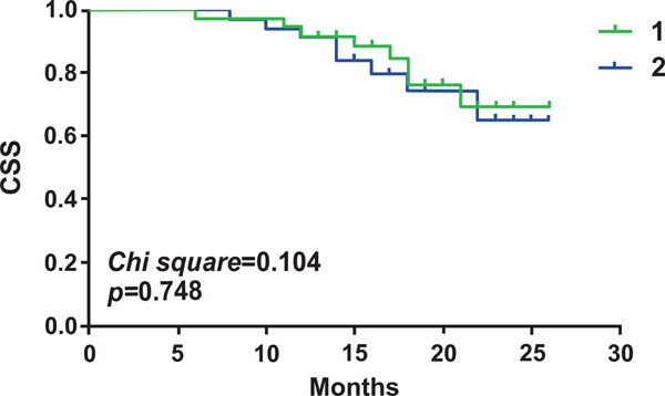
There was no significant difference in the survival time between two treatment methods (Fig. 1). We use all of the time periods for this analysis.

The survival time between a porous tantalum rod implant (1) and core decompression using multiple small drill holes (2) for the treatment of early femoral head necrosis. No significant difference in the survival time between two treatment methods.

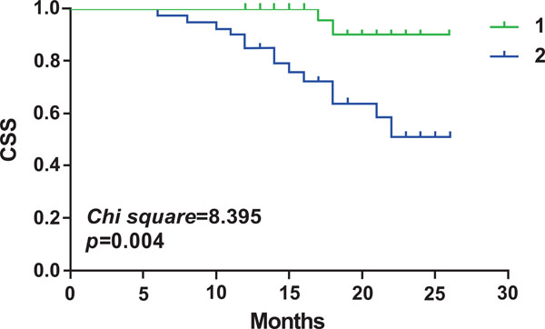
The survival time between stage I (1) and stage II (2) hips. Survival time is significantly shorter in stage II hips.

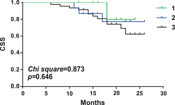
The survival time among osteonecrosis patients from different etiologies. (1) alcohol, (2) idiopathic, and (3) steroid. No significant difference in survival time among osteonecrosis patients from different etiologies.
Treatment Results in Different Stages
Preoperative and postoperative Harris hip scores were compared, there was a significant difference in Harris hip scores between stage I and stage II hips (p=0.017). There was a significant difference in the survival time between stage I and stage II hips (p=0.021). A porous tantalum rod implant and core decompression using multiple small drill holes for the treatment of early femoral head necrosis were better for stage I hips (Fig. 2).
Treatment Results with Different Etiologies
There was no statistical difference in Harris hip score improvement among osteonecrosis patients from different etiologies. There was no significant difference in survival time among osteonecrosis patients from different etiologies (p>0.05) (Fig. 3).
DISSCUSSION
The aim of this study was to evaluate the effect of a porous tantalum rod implant versus core decompression using small drill holes for the treatment of early femoral head necrosis. The clinical symptoms of the early stage patients improved according to the HHS by these two treatments. The average surgical time was 40 min, and the average volume of blood loss was 30 ml in both tow surgical groups. The average of Harris score was improved from 56.2 ± 7.1 preoperative to 80.2 ± 11.4 at the last follow-up in the rod implanted group (p < 0.05). The mean Harris score improved from 53.8 ± 6.6 preoperative to 79.7 ± 13.2 at the last follow-up in the small drill holes group (p<0. 05). No significant difference was observed in Harris score between two groups. At the last follow-up 28 of 36 hips were at the same radiographic stages as pre-operation, and 8 deteriorated in the rod implanted group. 26 of 34 hips were at the same radiographic stage as pre-operation, and 8 deteriorated in the small drill holes group. No significant difference was observed in radiographic stage between two groups. There was no favourable result on the outcome of a tantalum intervention implant compared to multiple small drill holes. A tantalum rod intervention and core decompression using multiple small drill holes were effective on the stage I hips.
CD is supposed to reduce the oedema-related intraosseous pressure so as to relieve pain [6]. It is reported that CD could contribute to the blood reperfusion, possibly associated with revascularisation and bone regeneration of the necrotic area [7]. It is reported that CD using multiple 3.2-mm drill holes allowed partial postoperative weight-bearing such as walking and climbing stairs. Only in cases of stumbling, the ultimate stress exists between 78 MPa and 150 MPa which was four times greater than during normal walking could induce a fracture [8-10]. CD through multiple small drill holes was superior to the tantalum implant for long-term evaluation because of the complete replenishment of the drill holes with new bone and the ingrowth behaviour of tantalum implant is still controversial for the finite element analysis which does not confirm complete bony ingrowth presumption. Furthermore, after the tantalum implantation, the MRI showed that a slight seam of fluid could be detected surrounding the implant which indicated that it was not complete bone ingrowth [3].
Porous tantalum which is also called trabecular metal has been used for a variety of surgical applications, such as hip and knee arthroplasty and bone graft substitute because of its excellent mechanical strength, porosity and biocompatibility [11, 12]. However, studies reported that the complication of the tantalum implant was subtrochanteric fracture [13, 14]. It was reported that there was about 10% fracture after CD using a 10-mm drill [15, 16]. However, it was reported that CD using multiple small drill holes caused no fracture during the follow-up time [17].
In addition, the cost for the tantalum implant is relatively high, which has to be considered when choosing the surgical procedure. Another disadvantage of the tantalum rod is that it is a foreign body that, in case of a deep infection, may have to be removed. This would be associated with a high risk of fracture. A possible advantage of the treatment is the earlier postoperative load-bearing without increased risk of femoral neck fracture, allowing the patients to resume their daily routine sooner. However, CD via multiple small drill holes would also allow similar postoperative load-bearing and seems to result in similar or even better clinical outcome without the prolonged implantation of an expensive tantalum implant.
Since the porous tantalum is expensive, and thought to be a “buy-time” technique, with the trouble when treated for the THR. It is better to use the multiple small drill holes to treat the early osteonecrosis.
CONCLUSION
The tantalum intervention implant with CD did not show considerable results compared to multiple small drill holes. Two methods for the treatment of early femoral head necrosis were better on the stage I hips.
CONFLICT OF INTERTST
The authors confirm that this article content has no conflict of interest.
ACKNOWLEDGEMENTS
This design has been granted practical patent by Patent Bureau of People’s Republic of China (No: 201020525718. X). This work was supported by Medical and Health Key Project of Guangzhou City, No. 2007-ZDi-11, No. 2009-zdi-04; and Guangdong Provincial Natural Science Foundation of China, No. 10151022001000005,S2011010000910


