All published articles of this journal are available on ScienceDirect.

The PFNA® Augmented in Revision Surgery of Proximal Femur Fractures
Abstract
Objectives:
Modern implants for proximal femur fracture treatment have clearly improved clinical results. However, complications, including cut-out and loss of reduction, requiring revision surgery still occur. A major challenge in these cases is a loss of bone stock due to the existing implant, which is usually exacerbated by osteoporosis. A potential solution is the augmentation of implants, for example, of the femoral neck blade using bone cement.
Materials and Methods:
Ten patients (five loosening of femoral neck implant, two pseudarthrosis, two implant failures and one acute fracture) were included. The initial hardware was removed and a PFNA augmented was implanted. The perforated femoral neck blade was augmented using polymethyl methacrylate cement. Clinical and radiological follow-up was performed at a mean of 5.4 months (SD ±4.34). The main outcome parameters were fracture healing and implant-related complications.
Results:
Technical handling was uneventful in all cases. No cement leakage into the joint occurred in any of the cases. The mean amount of cement injected was 5.3 ml. The fracture healed during follow-up in all cases except two patients who died from causes unrelated to the procedure and prior to complete consolidation. Problem-free elective hardware removal of the PFNA augmented was performed in two cases.
Discussion:
The PFNA augmented is a potential implant for joint-preserving revision surgery in proximal femur fractures. The augmentation improves implant anchorage in the impaired bone stock. In this preliminary series, no negative biological side effects of the cement (i.e. osteonecrosis) were observed.
INTRODUCTION
The treatment of proximal femur fractures has improved due to new implant designs in recent years [1, 2]. However, postoperative complication rates resulting from the procedure of up to 10.7% are still reported [3]. Although severe complications, including cut-out or cut-through, could be reduced significantly, they remain serious risk factors in patients with a weak bone stock [4-6]. In particular, the eccentric femoral neck implant position is often the main reason. It increases stress on the implant and thus failure of the implant bone anchorage [7]. In cases with complications, treatment is often challenging and technically demanding because of the diminished bone stock [8]. Therefore, particularly in the elderly, prosthetic joint replacement is frequently required, with all its accompanying intra- and postoperative risks, and is a form of major surgery in the geriatric patient population [9, 10]. Nevertheless, in cases which present with a destruction of the joint because of cut-through or cut-out, hip arthroplasty remains the only treatment option. However, in an age-related intact femoral joint or an increased perioperative risk due to co-morbidities, a joint-preserving and/or less-invasive procedure is preferable.
The PFNA augmented (Proximal Femoral Nail Antirotation, Synthes Inc., Oberdorf, Switzerland) displayed a significantly greater stability in biomechanical in vitro testing [11, 12]. Furthermore, the results ofthe primary treatment of osteoporotic proximal femur fractures are promising [13]. From a biomechanical perspective, these results are attributed to the enlargement of the bone-implant interface using cement. However, no clinical data is currently available regarding the suitability of the PFNA augmented in revision surgery of proximal femur fractures. The present study describes the surgical technique and early results in a preliminary case series of this method.
MATERIALS AND METHODS
Ten patients with a mean age of 76.5 years (SD±13.2) were treated using the PFNA augmented (Synthes Inc). In five cases revision surgery was performed because of a loosening of the femoral neck implant, in two cases because of a pseudarthrosis, in two cases because of an implant failure and in one case because of a new fracture with the implant being in situ (summarized in Table 1). In all the cases, the complete or respective part of the former implant was removed and a PFNA augmented was implanted. The insertion technique is the same as for the non-augmented PFNA, except for the perforated blade, which is shown in Fig. (1). To prevent cement leakage into the joint, a leakage test using contrast fluid was performed after insertion of the blade and prior to the injection of the cement. The
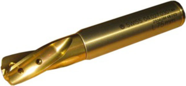
The perforated blade of the PFNA. Because of the perforation holes, the PMMA can cover the tip of the blade, which leads to a larger bone-implant interface.
augmentation cannula, shown in Fig. (2), was inserted through the aiming device. Under visualization using fluoroscopy, the contrast fluid (syringe with a luer lock) was administered and its distribution was monitored (Fig. 2). The augmentation was only performed after a leakage into the joint was ruled out by this procedure. Then the perforated helical-shaped femoral neck blade was augmented with polymethylmethacrylate (PMMA) cement (Traumacem V+, Synthes GmbH, Switzerland). The cement syringe connects to the augmentation cannula via a luer lock. The cannula has a side opening (red circle in Fig. 2), which is indicated at the handle, and thus the cement flow direction can be controlled. A recent study by Sermon et al. reported that the greatest improvement in stability was achieved with an augmentation cranial to the blade and at the tip of the blade [14]. The amount of injected cement was in accordance with the defect in the bone stock resulting from the former osteosynthesis and was monitored intraoperatively using fluoroscopy.
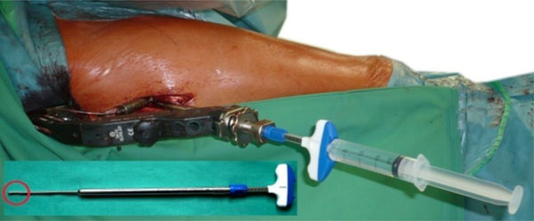
The augmentation cannula in a live setting (contrast fluid). The red circle shows the side opening of the cannula to direct the cement.
In Fig. (4), for example, the filling of the bone defect caused by the former osteosynthesis with bone cement is demonstrated. The mean follow-up time was 5.4 months (SD ± 4.34) and was performed clinically and using x-ray. The main outcome parameters were healing of the fracture and the appearance of implant failure.
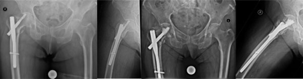
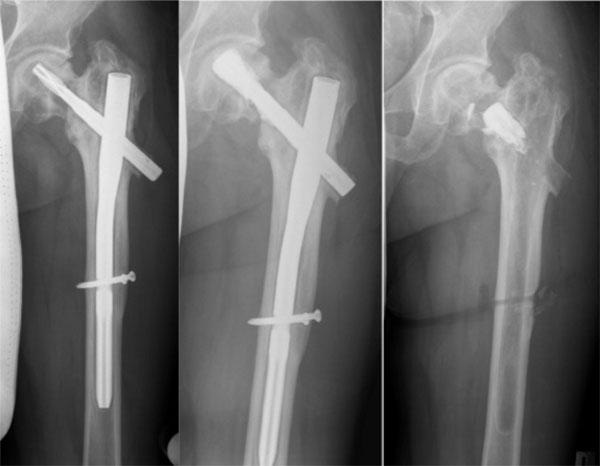
Patient no. 7. From left to right: Loosening of the femoral neck device 2 weeks after primary surgery. Implant removal and implantation of the PFNA augmented. Consolidation of the fracture 8 months postoperative.
RESULTS
PFNA augmented implantation was possible in all cases. Because the implantation technique of the implant itself does not differ from the non-augmented PFNA, no technical difficulties were observed. The augmentation process itself is an easy procedure which did not extensively extend the operation time. The duration ofthe leakage test and augmentation was approximately 4.7 min (SD±1.3). No cement leakage into the joint in any of the cases was observed. The mean amount of cement used was 5.3 ml (SD±2.7). Two patients died of natural causes during the follow up before healing of the fracture had occurred. In the remaining eight cases, the fracture healed uneventfully during the follow up. Figs. (3-5) show x-rays of patients no. 3, 7 and 10, respectively. In each case, the preoperative situation and the postoperative result are shown. In Fig. (5), the x-ray after implant removal is also presented. The implant removal of the augmented PFNA blade was comparable to the non-augmented version regarding surgical technique and duration of surgery. No implant-related complications were observed during the follow-up period. In two cases, the PFNA augmented was removed after consolidation of the fracture and because the patient requested it. An overview of the patients is provided in Table 1.
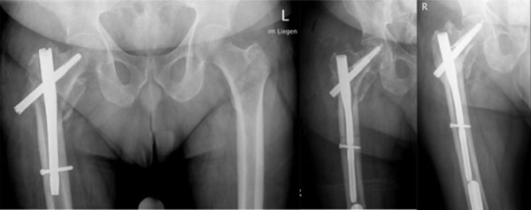
Patient no. 3. Cut-out of the femoral neck blade because of incorrect implant positioning (on the left). On the right, postoperative xrays after implant removal and implantation of the PFNA augmented are shown. The distribution of the cement in the bone defect is visible. The fracture healed after 3 months. There were no secondary complications after 5 months.
Patient no. 10 with loosening of the PFNA blade. Good visualization of the bone defect in the left image. In the middle, the postoperative x-ray after augmentation of the blade is shown. A filling of the defect can be seen. On the right is the x-ray after consolidation and implant removal after 17 months.
Summary of the patients’ data; f = female, m = male, DHS = Dynamic Hip Screw, PFNA = Proximal Femoral Nail Antirotation.
| No. | Age | Sex | Initial Treatment | Cause for Revision | Procedure | Follow-Up Time | Complications/ Comments | Healing Achieved |
|---|---|---|---|---|---|---|---|---|
| 1 | 70 | f | DHS | Implant failure | Implant removal and PFNA augmented | 7 months | Yes | |
| 2 | 79 | m | PFN | Loosening of the blade | Renewal of the blade and augmentation | 4 months | Yes | |
| 3 | 81 | f | PFN | Near cut-out due to malpositioning | Implant removal and PFNA augmented | 5 months | Yes | |
| 4 | 90 | f | PFNA | Peri-PFNA-fracture | Implant removal and PFNA long augmented | 6 weeks | Patient died 6 weeks later due to heart failure. No complications up to this point. | Unknown |
| 5 | 76 | f | PFNA | Implant failure (nail breakage) | Implant removal and PFNA long augmented | 3 months | Yes | |
| 6 | 91 | f | PFNA | Re-trauma with near cut-out | Implant removal and PFNA augmented | 4 weeks | Patient died 4 weeks later due to heart failure. No complications up to this point. | Unknown |
| 7 | 61 | m | Gamma Nail | Loosening of the femoral neck screw | Implant removal and PFNA augmented | 6 months | Yes | |
| 8 | 89 | f | PFNA | Re-trauma with dislocation of the blade | Implant removal and PFNA augmented | 3 months | Yes | |
| 9 | 78 | f | PFN | Pseudarthrosis | Implant removal + reaming and PFNA augmented | 7 months | Implant removal after 7 months | Yes |
| 10 | 50 | f | PFNA | Pseudarthrosis and loosening of the blade | Change of blade and augmentation | 17 months | Implant removal after 17 months | Yes |
DISCUSSION
The findings of the biomechanical testing regarding cement augmentation of the helical-shaped femoral neck blade show an improved anchorage in both foam as cadaveric models [11, 12]. Erhart et al. in a biomechanical in vitro study tested the augmented PFNA as a salvage procedure for lateral migration of the blade in a revision setting. After positioning of a helical blade in the femoral head, extraction and repositioning with augmentation pull-out strength and rotational stability were compared to the contra-lateral side, where a normal blade (non-augmented) was inserted. In this study, the revised blade with augmentation showed no inferiority in pull-out strength compared to the opposite side. The rotational stability was even greater [15]. Although pull-out strength and rotational stability do not fully cover the complex biomechanical situation in the proximal femur, the results do provide a good indication of the biomechanical performance of this implant.
A multicenter study by Kammerlander et al. with a mean follow-up time of 15 months showed no negative effects of augmentation, for example, cartilage damage or osteonecrosis [13]. Similarly, in our small series, such adverse events due to augmentation were not observed. Nevertheless, this procedure is not suitable for all patients. In particular, the destruction of the articular surface, as seen in
cut-out and cut-through, is an indication for prosthetic replacement or a contraindication for augmentation. Careful preoperative assessment of the clinical status of the patient and the x-ray is mandatory, as in any revision surgery. Our study is the first to report the use of the augmented PFNA not as a primary procedure, as shown by Kammerlander et al., but rather as an implant for revision in proximal femur fractures. Our experience with this implant shows promising results as a possible salvage procedure in challenging cases. Regardless of this aspect, a correct fracture reduction and implantation technique, particularly regarding the position of the femoral neck device, is mandatory. The results of our study show that even after augmenting the PFNA, hardware removal is still possible, as seen in the two respective cases. In Fig. (5), the postoperative x-ray shows that the cement had retreated into the cavity before the cement-blade interface broke. This indicates that in this patient, no strong osseous-cement interface could be established because of the existing defect. Because of the new cavity, a possible loss in the stability was suspected. Therefore, the patient was only allowed partial weight bearing for 6 weeks. After this period, full weight bearing was allowed. No secondary complications occurred.
Although promising, the results of this study are only preliminary because of the limited number of patients. Further studies are required to confirm our findings.
CONCLUSION
The PFNA augmented is a safe implant, which can be used in revision surgery in proximal femur fractures as a salvage procedure to avoid prosthetic replacement. Because the implantation technique does not differ (except for the perforated blade) from the established non-augmented PFNA, it is implemented easily. The augmentation procedure is safe and non-time consuming, and is easy to learn. Nevertheless, in the case of cut-out or medial implant protrusion with destruction of the joint, the proposed salvage technique is not feasible.
CONFLICT OF INTEREST
The authors confirm that this article content has no conflict of interest.
ACKNOWLEDGEMENTS
Declared none.

