REVIEW ARTICLE


MR Imaging of SLAP Lesions


Robert D. Boutin1, Richard A. Marder2, *
1 Department of Radiology, UC Davis School of Medicine, 4860 Y St., Suite 3100, Sacramento, CA 95817, USA
2 Department of Orthopaedic Surgery, UC Davis School of Medicine, 4860 Y St., Suite 3800, Sacramento, CA 95817, USA


Article Metrics

CrossRef Citations:
8
Total Statistics:

Full-Text HTML Views: 9136
Abstract HTML Views: 361
PDF Downloads: 670
ePub Downloads: 181
Total Views/Downloads: 10348
Unique Statistics:

Full-Text HTML Views: 5079
Abstract HTML Views: 247
PDF Downloads: 526
ePub Downloads: 146
Total Views/Downloads: 5998


Creative Commons License
© 2018 Boutin and Marder.

open-access license: This is an open access article distributed under the terms of the Creative Commons Attribution 4.0 International Public License (CC-BY 4.0), a copy of which is available at: (https://creativecommons.org/licenses/by/4.0/legalcode). This license permits unrestricted use, distribution, and reproduction in any medium, provided the original author and source are credited.

* Address correspondence to this author at the Department of Orthopaedic Surgery, UC Davis School of Medicine, 4860 Y St., Suite 3800, Sacramento, CA 95817 USA; Tel: 916-734-5566; E-mail: ramarder@ucdavis.edu


Abstract

Background:

SLAP lesions of the shoulder are challenging to diagnose by clinical means alone. Interpretation of MR images requires knowledge of the normal appearance of the labrum, its anatomical variants, and the characteristic patterns of SLAP lesions. In general, high signal extending anterior and posterior to the biceps anchor is the hallmark of SLAP lesions. Common diagnostic criteria for a SLAP lesion by MR or MR arthrography include the following: presence of a laterally curved, high signal intensity in the labrum on a coronal image, multiple or branching lines of high signal intensity in the superior labrum on a coronal image, full-thickness detachment with irregularly marginated high signal intensity and/or separation >2 mm on conventional MRI or 3 mm on MR arthrography between the labrum and glenoid on a coronal image, and a paralabral cyst extending from the superior labrum.

Methods:

MR diagnosis of SLAP tears may be improved with provocative maneuvers, such as longitudinal traction of the arm or positioning of the shoulder in abduction and external rotation during imaging. The use of intra-articular contrast distends the joint similar to what occurs during arthroscopy and forced diffusion under the labrum may improve the ability to detect SLAP lesions that might not be seen with standard MR. Improved diagnostic accuracy for SLAP tears is seen with 3-T compared with 1.5-T MR imaging, with or without intra-articular contrast material.

Conclusion:

Regardless of MR findings, however, physicians should be cautious when recommending surgery in the patient with a vague clinical picture. The patient’s history, physical exam, and imaging evaluation all should be considered together in making the decision to proceed with surgery.

Keywords: SLAP tear, MRI, Superior labrum, Biceps anchor, MR arthrography, Intra-articular.


1. INTRODUCTION

SLAP lesions of the shoulder are challenging to diagnose by clinical means alone [1]. Injury occurs from inferior traction on the shoulder, as well as excessive tension on and twisting of the Long Head of Biceps tendon (LHB) at its insertion along the superior labrum that occurs during the cocking motion of throwing [2, 3]. These mechanisms of injury are also associated with a myriad of other shoulder conditions including rotator cuff tendinopathy, bicipital tendinopathy and subluxation, internal impingement, and glenohumeral instability. Thus, history, while helpful, only suggests the diagnosis of a SLAP lesion.

Standard shoulder physical examination of the shoulder may be normal unless instability is present. Specialized exam tests have been developed that help the LHB insertion to assist in the clinical diagnosis. Such provocative maneuvers include the active compression test, biceps load test II, Speed’s test, the resisted supination external rotation test, and the dynamic labral shear test [4-7]. Variable success in predicting a SLAP lesion has been reported with these tests in systematic reviews [8, 9], and one rigorous Level I study has shown poor correlation with arthroscopy [10]. When used in combination, the accuracy to diagnosis a SLAP lesion by physical exam may be improved [11-13].

Correct diagnosis of SLAP lesions is important because the ability to confidently predict that a patient does not have a SLAP lesion can prevent an arthroscopy that simply discovers a normal superior labrum. Furthermore, the ability to accurately diagnose the type of SLAP lesion allows pre-operative planning, for not only the surgical procedure, but also post-operative rehabilitation, which can be prolonged in certain cases. Imaging is a potentially helpful tool in diagnosing a SLAP tear pre-operatively. Both MR and MR arthrography have been used with varying degrees of success [14-18].

2. IMAGING

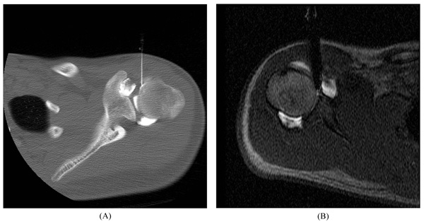
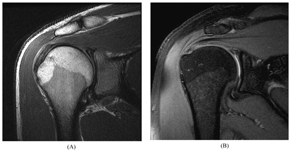
At our institution, routine MR of the shoulder is performed using a 1.5–Tesla (T) or 3-T scanner to obtain Fast Spin-Echo (FSE) proton-density and fat-suppressed T2-weighted images in the axial, coronal oblique, and sagittal oblique planes. Images in the axial and coronal oblique planes are most useful for the evaluation of SLAP lesions. For MR arthrography, percutaneous injection of diluted gadolinium (12 mL, 1:200 dilution) into the glenohumeral joint is performed using an anterior approach. Radiologists usually confirm intra-articular injections fluoroscopically, although sonography, CT, or MRI also may be used (Fig. 1). MR scanning begins within 30 minutes of injection, and commonly includes FSE fat-suppressed T1-weighted images in all three planes (which highlight any contrast material filling labral tear), as well as FSE coronal fat-suppressed T2-weighted images (which show any paralabral cyst) and sagittal T1-weighted images (which show fatty muscle atrophy associated with a chronic cuff tear or muscle denervation). Detailed, updated standard of practice imaging protocols are posted online [19].

Fig. (1). Arthrogram procedure. Anterior approach of needle for image-guided glenohumeral joint injection of contrast material using (A) CT and (B) MRI.

3. IMAGING INTERPRETATION

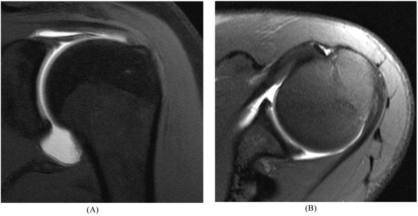
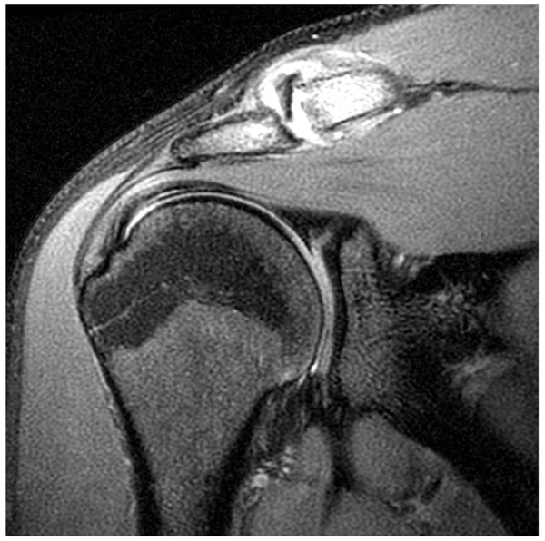
Correct interpretation of MR images requires knowledge of the normal appearance of the labrum, its anatomical variants, and the characteristic patterns of SLAP lesions [20]. The fibrocartilaginous labrum most often has a triangular structure, but it may change shape dynamically with traction from the capsule or glenohumeral ligaments (e.g., appear round or flattened). Regardless, if there is no high signal either at the labral–chondral junction or through the labrum, the labrum should be graded as normal [21] (Fig. 2).

Fig. (2). Normal labrum. 14 year-old male athlete with an unequivocally normal labrum. (A) Coronal and (B) axial fat-suppressed T1-weighted MR arthrogram images show normal low signal intensity fibrocartilage; no contrast material extends into the labrum or into the chondrolabral junction.

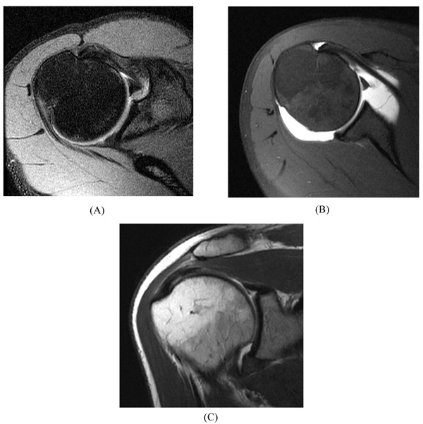
Anatomic variations in the labrum most commonly occur in the anterior-superior quadrant (between 12 and 3 o’clock), including the Buford complex, sublabral sulcus, and sublabral foramen [22-25]. A Buford complex (Fig. 3) is a distinct congenital condition that occurs in approximately 1 to 7% of normal shoulders [25]. This anatomic variant is defined by an absent upper anterior quadrant of the labrum and a thickened, cord-like middle glenohumeral ligament that inserts directly into the superior labrum. Interestingly, although the Buford complex is regarded as an anatomic variant, recent literature indicates that it may predispose patients to the development of SLAP lesions [26], perhaps due to altered biomechanics [27].

The sublabral sulcus (also referred to as a sublabral recess) and sublabral foramen are frequent anatomic variations in which a segment of the labrum is not attached to the underlying glenoid. The sublabral sulcus is particularly important; a partially unattached labrum with some form of a superior recess is reported in up to 75% of patients without a superior labral tear [28]. This occurs at the apex of the superior labrum, but classically does not extend posteriorly to the LHB attachment. The depth of the recess is variable, but typically less than 2 mm [25].

Beginning with the classic work by DePalma [29], authors have suggested that the detachment at the chondrolabral junction is an age-dependent degenerative phenomenon because superior recesses are not present at birth, and the incidence and depth of recesses increases with age [29-31]. Indeed, the high prevalence of superior chondrolabral detachment and marginal benefits of SLAP repair after patients reach the age of 40 to 45 years should be considered when making treatment decisions [27, 32]. Although, a sublabral sulcus typically appears as a cleft of smooth, medially curved increased signal at the chondrolabral junction that does not extend posterior to the biceps anchor, these normal variants reportedly can extend posterior to the biceps anchor and therefore may be misinterpreted as a labral tear [29]. Despite that, it must be emphasized that high signal extending anterior and posterior to the biceps anchor is the hallmark of SLAP lesions.

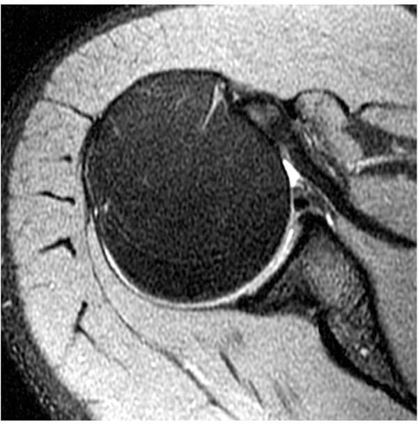
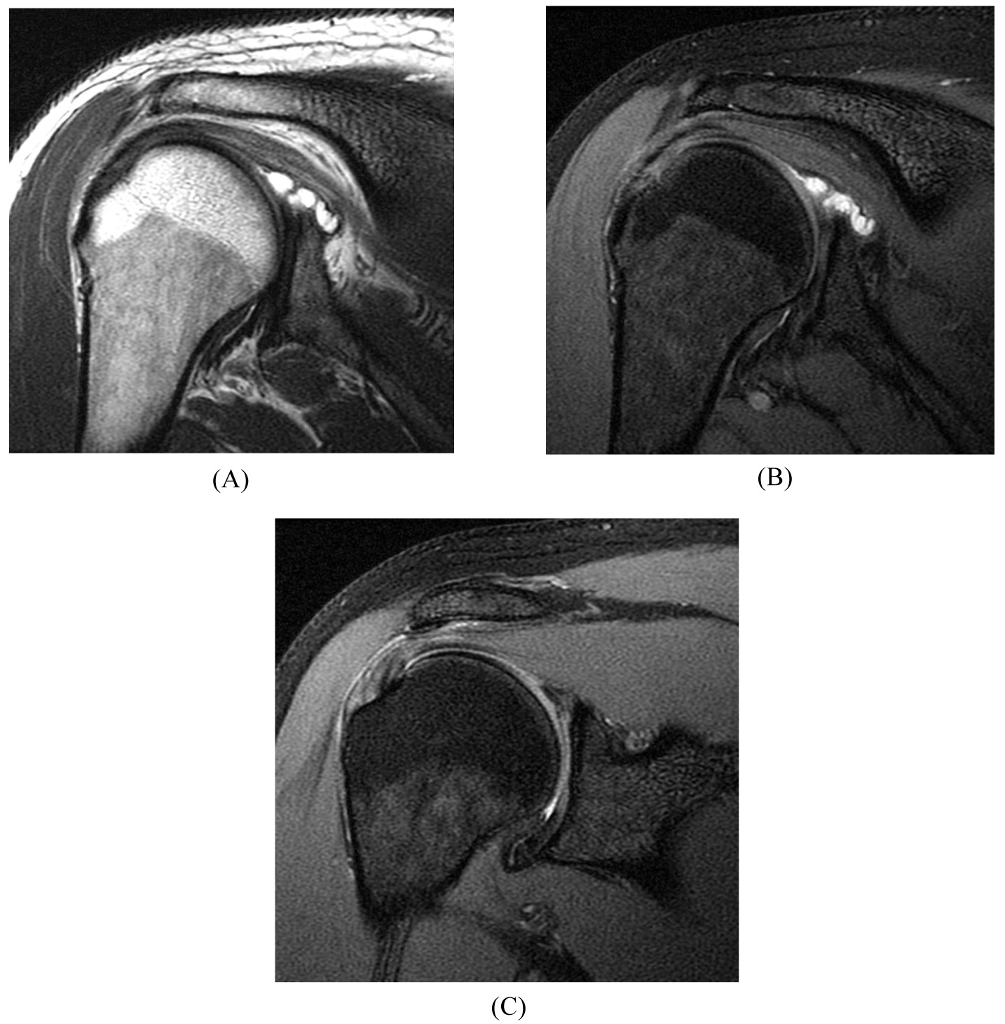
The sublabral foramen occurs only in the upper anterior quadrant of the glenoid and does not involve the LHB insertion (Fig. 4). The sublabral foramen appears as a smoothly marginated separation of the anterosuperior labrum from the underlying glenoid (over a range from approximately 3 to 11 mm cranial to caudal) and has a prevalence of approximately 11% [23].

Fig. (3). Buford complex. (A) Axial fat-suppressed T2-weighted image of a 39 year-old male athlete with an absent anterosuperior labrum and a thickened, cord-like middle glenohumeral ligament. (B) Axial fat-suppressed T1-weighted MR arthrogram image in a 44 year-old orthopaedist, a female athlete with a Buford complex. (C) Coronal T1-weighted MR arthrogram image in the same shoulder shows a superior labral tear.

Fig. (4). Sublabral foramen. (A) Axial fat-suppressed T2-weighted image of a 29 year-old male athlete with smoothly marginated, full-thickness separation of the labrum isolated to the anterosuperior quadrant.

Diagnostic criteria for a SLAP lesion by MR or MR arthrography most commonly include the following [29, 33]:

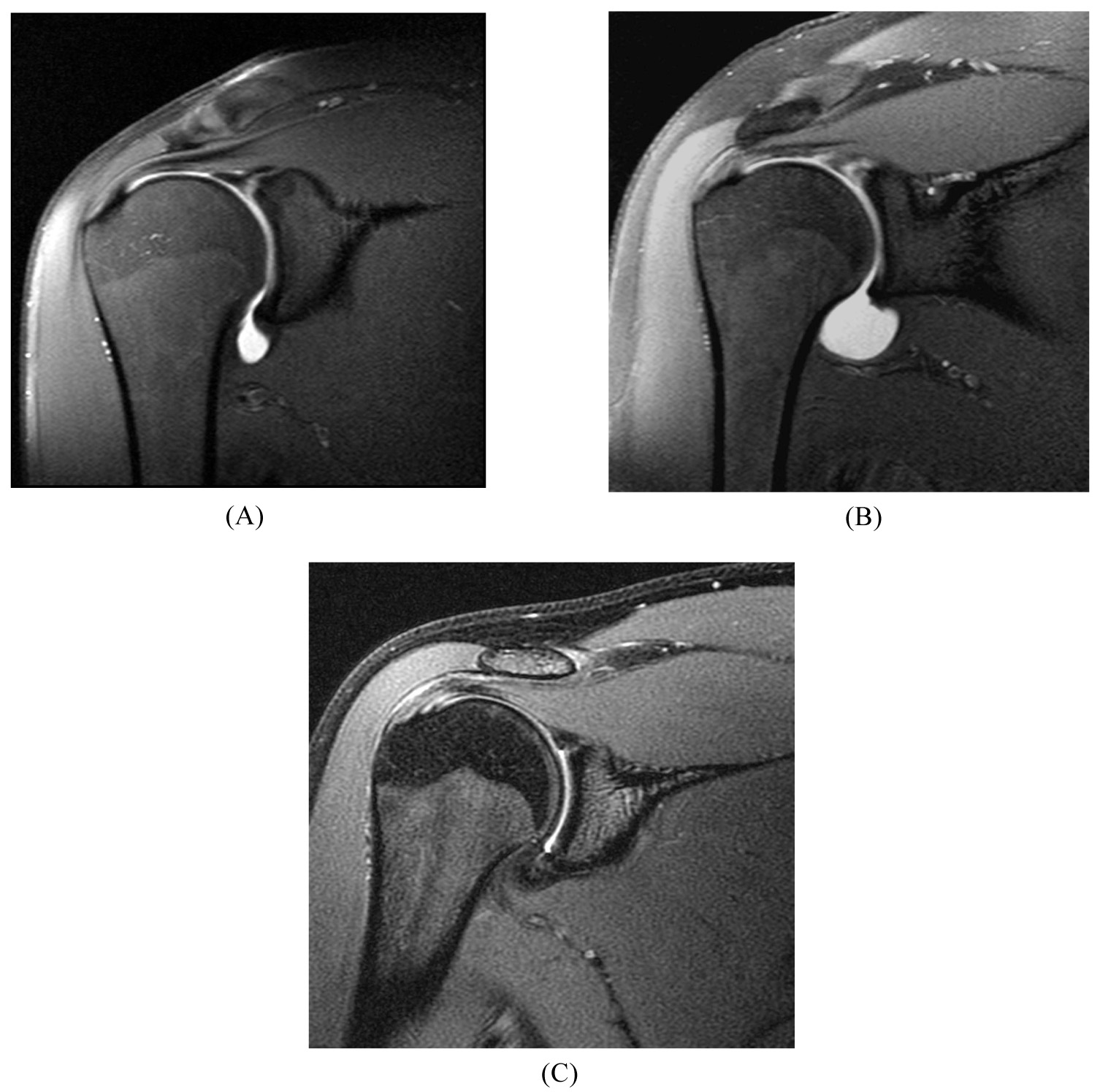
  1. laterally curved, linear signal in the labrum on coronal imaging (Fig. 5);
  2. multiple or branching lines of high signal intensity in the superior labrum on coronal imaging (Fig. 6);
  3. full-thickness detachment with irregularly marginated high signal intensity and/or wide separation (e.g., width >2 mm on conventional MRI or 3 mm on MR arthrography) between the labrum and glenoid on coronal imaging (Fig. 7);
  4. a paralabral cyst extending from the superior labrum [34] (Fig. 8).
Fig. (5). SLAP tear. Laterally curved linear signal located posterior to the biceps anchor in (A) a 22 year-old collegiate baseball pitcher, on a coronal intermediate-weighted image and (B) a 27 year-old overhead athlete, on a coronal fat-suppressed T2-weighted image.

Fig. (6). SLAP tear. Branching linear signal located posterior to the biceps anchor in a 19 year-old collegiate athlete on a coronal fat-suppressed T2-weighted image. Also note the presence of distal clavicle erosion and edema in this athlete with distal clavicular osteolysis owing to weight lifting.

Fig. (7). SLAP tear. Full-thickness detachment with linear signal separating the superior labrum from the glenoid rim in (A) a 21 year-old collegiate thrower, on a coronal fat-suppressed T1-weighted MR arthrogram, (B) a 28 year-old female athlete, on a coronal fat-suppressed T1-weighted MR arthrogram, and (C) a 19 year-old male athlete, on a coronal fat-suppressed T2-weighted MR image (without arthrography).

Additional MR diagnostic criteria also have been proposed. The “sulcus score” has been developed to help differentiate between a superior labral sulcus versus a type II SLAP lesion [35]. The sulcus score quantifies the overall magnitude of the sulcus by accounting for both the sulcus depth and length. (The sulcus score is calculated by multiplying the depth (“grade”) of the sulcus by the length of the sulcus (the number of “hours” around the “clock” of the glenoid).

4. DISCUSSION

MR and MR arthrography with 1.5-T and 3-T scanners are currently the mainstays of imaging used to diagnose SLAP lesions prior to arthroscopy. Other modalities have been used to image the superior labrum, including CT arthrography which has been successfully utilized and ultrasound which has not been shown to be efficacious [36-38].

There is some evidence that the MR diagnosis of SLAP tears may be improved with provocative maneuvers, such as longitudinal traction of the arm [39] or positioning the shoulder in abduction and external rotation during imaging [40, 41]. The use of intra-articular contrast distends the joint similar to what occurs during arthroscopy and may improve the ability to detect SLAP lesions by filling any labral tear. Recent systematic reviews and meta-analyses conclude that MR arthrography is better than standard MR in detecting labral tears [42-44], with a higher sensitivity (80% vs. 63%) but similar specificity (91% vs. 87%) for SLAP tears [43].

Fig. (8). SLAP tear in a 39 year-old patient with a history of a negative c-spine MRI. Lobulated paralabral cyst at the posterosuperior glenoid rim due to a labral tear on (A) a coronal intermediate-weighted image and (B) a coronal fat-suppressed T2-weighted image. (C) At the adjacent superior labrum near the biceps anchor, there is subtle, laterally curved, linear signal in the labrum indicative of a SLAP tear (without arthrography).

Is there improved diagnostic accuracy with 3-T MR imaging? Although there is substantial heterogeneity in published studies and further research would be appropriate, the most recent pooled data suggests that improved diagnostic accuracy for SLAP tears results with 3-T compared with 1.5-T MR imaging [43, 44], with or without intra-articular contrast material (sensitivity, 78%–84% vs 79%–81% and specificity, 95%–99% vs 67%–84%) [43]. The U.S. Food and Drug Administration recently approved ultrahigh-field 7-T MR scanners for clinical use [45], but only preliminary studies have been performed on the shoulder [46].

CONCLUSION

Continued technological advancements may result in improved accuracy of MR diagnosis of SLAP lesions. Regardless of any MR findings, however, physicians should be cautious when recommending surgery in the patient with a vague clinical picture. The patient’s history, physical exam, and imaging evaluation all should be considered together in making the decision to proceed with surgery.

CONSENT FOR PUBLICATION

Not applicable.

CONFLICT OF INTEREST

The authors declare no conflict of interest, financial or otherwise.

ACKNOWLEDGEMENTS

Declared none.

REFERENCES

[1] Snyder SJ, Karzel RP, Del Pizzo W, Ferkel RD, Friedman MJ. SLAP lesions of the shoulder. Arthroscopy 1990; 6(4): 274-9.
[2] Burkhart SS, Morgan CD. The peel-back mechanism: Its role in producing and extending posterior type II SLAP lesions and its effect on SLAP repair rehabilitation. Arthroscopy 1998; 14(6): 637-40.
[3] Maffet MW, Gartsman GM, Moseley B. Superior labrum-biceps tendon complex lesions of the shoulder. Am J Sports Med 1995; 23(1): 93-8.
[4] Kim SH, Ha KI, Ahn JH, Kim SH, Choi HJ. Biceps load test II: A clinical test for SLAP lesions of the shoulder. Arthroscopy 2001; 17(2): 160-4.
[5] Myers TH, Zemanovic JR, Andrews JR. The resisted supination external rotation test: A new test for the diagnosis of superior labral anterior posterior lesions. Am J Sports Med 2005; 33(9): 1315-20.
[6] O’Brien SJ, Pagnani MJ, Fealy S, McGlynn SR, Wilson JB. The active compression test: A new and effective test for diagnosing labral tears and acromioclavicular joint abnormality. Am J Sports Med 1998; 26(5): 610-3.
[7] Sodha S, Srikumaran U, Choi K, Borade AU, McFarland EG. Clinical assessment of the dynamic labral shear test for superior labrum anterior and posterior lesions. Am J Sports Med 2017; 45(4): 775-81.
[8] Gismervik SØ, Drogset JO, Granviken F, Rø M, Leivseth G. Physical examination tests of the shoulder: A systematic review and meta-analysis of diagnostic test performance. BMC Musculoskelet Disord 2017; 18(1): 41.
[9] Hanchard NC, Lenza M, Handoll HH, Takwoingi Y. Physical tests for shoulder impingements and local lesions of bursa, tendon or labrum that may accompany impingement. Cochrane Database Syst Rev 2013; (4): CD007427.
[10] Cook C, Beaty S, Kissenberth MJ, Siffri P, Pill SG, Hawkins RJ. Diagnostic accuracy of five orthopedic clinical tests for diagnosis of superior labrum anterior posterior (SLAP) lesions. J Shoulder Elbow Surg 2012; 21(1): 13-22.
[11] Oh JH, Kim JY, Kim WS, Gong HS, Lee JH. The evaluation of various physical examinations for the diagnosis of type II superior labrum anterior and posterior lesion. Am J Sports Med 2008; 36(2): 353-9.
[12] Hegedus EJ, Goode AP, Cook CE, et al. Which physical examination tests provide clinicians with the most value when examining the shoulder? Update of a systematic review with meta-analysis of individual tests. Br J Sports Med 2012; 46(14): 964-78.
[13] Somerville LE, Willits K, Johnson AM, et al. Clinical assessment of physical examination maneuvers for superior labral anterior to posterior lesions. Surg J (N Y) 2017; 3(4): e154-62.
[14] Amin MF, Youssef AO. The diagnostic value of magnetic resonance arthrography of the shoulder in detection and grading of SLAP lesions: comparison with arthroscopic findings. Eur J Radiol 2012; 81(9): 2343-7.
[15] Connell DA, Potter HG, Wickiewicz TL, Altchek DW, Warren RF. Noncontrast magnetic resonance imaging of superior labral lesions. 102 cases confirmed at arthroscopic surgery. Am J Sports Med 1999; 27(2): 208-13.
[16] Phillips JC, Cook C, Beaty S, Kissenberth MJ, Siffri P, Hawkins RJ. Validity of noncontrast magnetic resonance imaging in diagnosing superior labrum anterior-posterior tears. J Shoulder Elbow Surg 2013; 22(1): 3-8.
[17] Waldt S, Burkart A, Lange P, Imhoff AB, Rummeny EJ, Woertler K. Diagnostic performance of MR arthrography in the assessment of superior labral anteroposterior lesions of the shoulder. AJR Am J Roentgenol 2004; 182(5): 1271-8.
[18] Sheridan K, Kreulen C, Kim S, Mak W, Lewis K, Marder R. Accuracy of magnetic resonance imaging to diagnose superior labrum anterior-posterior tears. Knee Surg Sports Traumatol Arthrosc 2015; 23(9): 2645-50.
[19] Musculoskeletal Imaging and Intervention Section: Imaging Protocols and Scanning Parameters University of Wisconsin Department of Radiology 2017. Available from: https://www.radiology.wisc.edu/sections/msk/protocols.php
[20] Herzog RJ, Potter HG. Letter to the Editor: What does a shoulder mri cost the consumer? Clin Orthop Relat Res 2017; 475(6): 1749-50.
[21] Tuite MJ, Currie JW, Orwin JF, Baer GS, del Rio AM. Sublabral clefts and recesses in the anterior, inferior, and posterior glenoid labrum at MR arthrography. Skeletal Radiol 2013; 42(3): 353-62.
[22] Smith DK, Chopp TM, Aufdemorte TB, Witkowski EG, Jones RC. Sublabral recess of the superior glenoid labrum: study of cadavers with conventional nonenhanced MR imaging, MR arthrography, anatomic dissection, and limited histologic examination. Radiology 1996; 201(1): 251-6.
[23] Stoller DW. MR arthrography of the glenohumeral joint. Radiol Clin North Am 1997; 35(1): 97-116.
[24] Williams MM, Snyder SJ, Buford D Jr. The Buford complex--the “cord-like” middle glenohumeral ligament and absent anterosuperior labrum complex: A normal anatomic capsulolabral variant. Arthroscopy 1994; 10(3): 241-7.
[25] Itoigawa Y, Itoi E. Anatomy of the capsulolabral complex and rotator interval related to glenohumeral instability. Knee Surg Sports Traumatol Arthrosc 2016; 24(2): 343-9.
[26] Knesek M, Skendzel JG, Dines JS, Altchek DW, Allen AA, Bedi A. Diagnosis and management of superior labral anterior posterior tears in throwing athletes. Am J Sports Med 2013; 41(2): 444-60.
[27] Kanatli U, Ozturk BY, Bolukbasi S. Anatomical variations of the anterosuperior labrum: Prevalence and association with type II superior labrum anterior-posterior (SLAP) lesions. J Shoulder Elbow Surg 2010; 19(8): 1199-203.
[28] Gustas CN, Tuite MJ. Imaging update on the glenoid labrum: Variants versus tears. Semin Musculoskelet Radiol 2014; 18(4): 365-73.
[29] DePalma AF. The classic. Recurrent dislocation of the shoulder joint. Ann Surg. 1950;132:1052-1065. Clin Orthop Relat Res 2008; 466(3): 520-30.
[30] Koziak A, Chuang MJ, Jancosko JJ, Burnett KR, Nottage WM. Magnetic resonance arthrography assessment of the superior labrum using the BLC system: Age-related changes mimicking SLAP-2 lesions. Skeletal Radiol 2014; 43(8): 1065-70.
[31] Pfahler M, Haraida S, Schulz C, et al. Age-related changes of the glenoid labrum in normal shoulders. J Shoulder Elbow Surg 2003; 12(1): 40-52.
[32] Schwartzberg R, Reuss BL, Burkhart BG, Butterfield M, Wu JY, McLean KW. High prevalence of superior labral tears diagnosed by MRI in middle-aged patients with asymptomatic shoulders. Orthop J Sports Med 2016; 4(1): 2325967115623212.
[33] Jung JY, Jee WH, Park MY, Lee SY, Kim YS. SLAP tears: Diagnosis using 3-T shoulder MR arthrography with the 3D isotropic turbo spin-echo space sequence versus conventional 2D sequences. Eur Radiol 2013; 23(2): 487-95.
[34] Kessler MA, Stoffel K, Oswald A, Stutz G, Gaechter A. The SLAP lesion as a reason for glenolabral cysts: A report of five cases and review of the literature. Arch Orthop Trauma Surg 2007; 127(4): 287-92.
[35] Mihata T, McGarry MH, Tibone JE, Abe M, Lee TQ. Type II SLAP lesions: A new scoring system--the sulcus score. J Shoulder Elbow Surg 2005; 14(1)(Suppl. S): 19S-23S.
[36] Jarraya M, Roemer FW, Gale HI, Landreau P, D’Hooghe P, Guermazi A. MR-arthrography and CT-arthrography in sports-related glenolabral injuries: A matched descriptive illustration. Insights Imaging 2016; 7(2): 167-77.
[37] Choi BH, Kim NR, Moon SG, Park JY, Choi JW. Superior labral cleft after superior labral anterior-to-posterior tear repair: CT arthrographic features and correlation with clinical outcome. Radiology 2016; 278(2): 441-8.
[38] Pavic R, Margetic P, Bensic M, Brnadic RL. Diagnostic value of US, MR and MR arthrography in shoulder instability. Injury 2013; 44(Suppl. 3): S26-32.
[39] Chan KK, Muldoon KA, Yeh L, et al. Superior labral anteroposterior lesions: MR arthrography with arm traction. AJR Am J Roentgenol 1999; 173(4): 1117-22.
[40] Chhadia AM, Goldberg BA, Hutchinson MR. Abnormal translation in SLAP lesions on magnetic resonance imaging abducted externally rotated view. Arthroscopy 2010; 26(1): 19-25.
[41] Borrero CG, Casagranda BU, Towers JD, Bradley JP. Magnetic resonance appearance of posterosuperior labral peel back during humeral abduction and external rotation. Skeletal Radiol 2010; 39(1): 19-26.
[42] Smith TO, Drew BT, Toms AP. A meta-analysis of the diagnostic test accuracy of MRA and MRI for the detection of glenoid labral injury. Arch Orthop Trauma Surg 2012; 132(7): 905-19.
[43] Symanski JS, Subhas N, Babb J, Nicholson J, Gyftopoulos S. Diagnosis of superior labrum anterior-to-posterior tears by using MR imaging and MR arthrography: A systematic review and meta-analysis. Radiology 2017; 285(1): 101-13.
[44] Arirachakaran A, Boonard M, Chaijenkij K, Pituckanotai K, Prommahachai A, Kongtharvonskul J. A systematic review and meta-analysis of diagnostic test of MRA versus MRI for detection superior labrum anterior to posterior lesions type II-VII. Skeletal Radiol 2017; 46(2): 149-60.
[45] FDA News Release FDA clears first 7T magnetic resonance imaging device US Food and Drug Administration; Silver Spring, MD 2017. Available from: https://www.fda.gov/NewsEvents/Newsroom/PressAnnouncements/ucm580154.htm
[46] Rietsch SHG, Pfaffenrot V, Bitz AK, et al. An 8-channel transceiver 7-channel receive RF coil setup for high SNR ultrahigh-field MRI of the shoulder at 7T. Med Phys 2017; 44(12): 6195-208.